Volume 6 Number 3

Editorial
Honavar PU

Pseudopelvic Tenderness
Parulekar SV

Port Wine Stain In Pregnancy
Parulekar SV

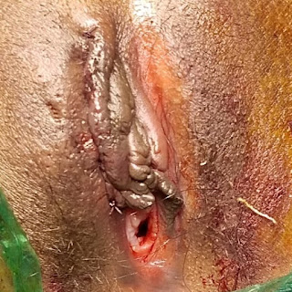
Recurrent Clitoral Cyst
Singhania N, Gaikwad C, Gupta AS.

Pemphigus Vulgaris In Pregnancy
Pradhan M, Vaidya A, Gupta AS.

Acute Pelvic Inflammatory Disease Mimicking Ruptured Ectopic Pregnancy
Kumar M, Shah R, Warke HS.

Genital Wart In Pregnancy
Jahan N, Tiwari N, Chaudhari HK.

Postoperative Lower Segment Cesarean Section Uterine Wound Dehiscence
Misar P, Samant P, Naykodi P.

Prolonged Bleeding After Coitus: Glanzmann Thromboasthenia
ŞahinBinel K, Yavasoglu I.


Remembering Past Greats: Robert Lawson Tait
Prasad M, Venkatesh S.


Editorial

Honavar PU

Greetings from the editors as we bring forth the third issue of this year. The month of February is observed as the ‘International Prenatal Infections Prevention’ month. This is to create worldwide awareness of the burden and impact of prenatal infections and to promote measures to improve maternal and neonatal outcome. Prenatal infections are infections that are transmitted to the fetus or newborn during the antenatal period or during delivery. Prenatal infections of obstetric and neonatal significance are viral infections like Human immunodeficiency virus (HIV), Hepatitis B, Hepatitis E, Toxoplasmosis, Rubella, Cytomegalovirus, Herpes, Parvovirus, Varicella Zoster, bacterial infections like Syphilis, Group B Streptococcus, Listeria, tropical diseases like malaria and chronic diseases like tuberculosis to list a few. Around 2 to 3% of congenital anomalies are accountable to prenatal infections. HIV is a sexually transmitted or blood borne virus that affects the T lymphocytes in the body leading to failure of immune system and eventually Acquired Immunodeficiency Syndrome (AIDS). Mother to child transmission is a cause of infection in a newborn. Without treatment, rate of transmission to a newborn is around 15 to 45%. Screening and anti -retroviral treatment in pregnancy has reduced it to less than 5%. Hepatitis B infection can lead to acute or chronic hepatitis, liver cirrhosis or hepatocellular carcinoma. Infection in infancy can lead to high rate of persistent infection and mother to child transmission accounts for approximately 50% of chronic infection cases. Screening for maternal infection and neonatal immunization can reduce the risk of infectivity. Although the incidence of syphilis has reduced over the years, Congenital syphilis is still a cause of early fetal loss, stillbirths, prematurity, neonatal deaths or debilitating conditions like deafness and interstitial keratitis. Serological screening can pick up the infection. Toxoplasmosis, Rubella, Cytomegalovirus, Herpes (TORCH), parvovirus and varicella zoster infections are notorious in pregnancy. They may be asymptomatic or may present as a flu like illness in a mother but have consequences of spontaneous abortions, intrauterine fetal death, growth restriction, congenital anomalies, prematurity, still births or live births with mental retardation, developmental delay, microcephaly, hearing and visual defects. Clinical suspicion, peculiar features on ultrasonography with immunological tests can help in the diagnosis of these infections. Diagnosis, severity of affection and the gestational age can guide in the management of these pregnancies. Fetal medicine expertise can be sought for the same. Listeriosis is a food borne infection caused due to consumption of raw and unprocessed dairy and meat products. Though less prevalent in India, suspicion should be high as when noted can cause fatal fetal or neonatal complications in around 25% cases. Tropical diseases like malaria may have transplacental transmission of the malarial parasites leading to abortions, growth restriction, prematurity and fetal death. Diagnosis based on history and detection of malarial parasites in the peripheral smear or on histopathological examination of placenta and treatment with safe antimalarials is advocated. Another prevalent disease in India is tuberculosis. Vertical transmission is a known mode of transmission leading to growth restriction, low birth weight, failure to thrive and increased risk of neonatal mortality. Prompt diagnosis and accessible treatment through Revised National Tuberculosis Control program (RNTCP) can lead to a better outcome. Recently, awareness of Group B Streptococcal (GBS) infection has gained momentum in the wake of poor maternal and neonatal outcome in women found positive for the organism. Western countries have incorporated it as a part of their antenatal counseling. Most women are asymptomatic carriers. An untreated pregnancy would result in neonatal pneumonia, meningitis, sepsis and mortality. Infant mortality rate is as high as 55%. Intrapartum benzyl penicillin reduces the incidence of early onset GBS disease. Role of GBS is largely unrecognized and underestimated although India is amongst the first five countries in number of pregnant women to be colonized with the bacterium. Controversy exists regarding the ideal preventive strategy and need for universal antenatal screening. At least, women with neonates at the risk of GBS infection need attention. These would include those with preterm births, prolonged rupture of membranes, preterm prolonged rupture of membranes, intrapartum fever, any antenatal maternal culture of GBS or previously affected child. More evidence based studies from the Indian subcontinent can help in framing guidelines for local management. Numerous activities and awareness sessions are conducted across the globe for implementing better policies ultimately with the aim of improving maternal and neonatal wellbeing. Hope our readers are enlightened timely on these topics and help each one of us contribute to a better and healthier society.

Pseudopelvic Tenderness

Author Information

Parulekar SV
(Professor and Head, Department of Obstetrics and Gynecology, Seth G S Medical College & K E M Hospital, Mumbai, India.)

Abstract

Pelvic tenderness in a woman can be caused by a number of conditions, like acute pelvic infection, endometriosis and torsion of adnexal structures. It is conventionally evaluated by an algorithmic approach, the final investigation being a laparoscopy. We encounter some cases in which no cause is found after extensive evaluation, including a laparoscopy as the final diagnostic case. An innovative approach was used in one case recently, which helped avoid an unnecessary laparoscopy.

Introduction

Pelvic tenderness is caused by inflammation of pelvic structures, which may be an infection like acute salpingo-oophoritis, pelvic peritonitis and pelvic cellulitis, or endometriosis or acute torsion of normal adnexa or adnexal mass like an ovarian tumor, or acute hemorrhage as from an ectopic pregnancy or corpus luteum hematoma. The diagnosis can usually be made by history, clinical examination, laboratory tests, imaging and if required, a laparoscopy. Sometimes no cause can be found despite all tests being done. A simple test is described to differentiate a vaginal pain due to hyperesthesia, so that an unnecessary laparoscopy was avoided.

Case Report

A 45 year old woman, para 2 living 2, presented with a complaint of something coming out per vaginum. Her menstrual cycles were regular, every 28-30 days, mildly painful and with moderate flow for 3-4 days. Her sexual history were normal. There was no dyspareunia. Her medical and surgical history was not contributory. She had two normal vaginal deliveries in the past. Her general and systemic examination revealed no abnormality. Abdominal examination findings were normal. A speculum examination showed healthy cervix and vagina, a moderate rectocele of right lateral type and a lax perineum. A bimanual examination showed lateral forniceal tenderness, a normal sized retroverted uterus, and no pelvic masses. The sensation over the remaining vagina was normal. A diagnosis of a rectocele, perineal laxity and possibly pelvic inflammatory disease was made. She was prescribed, as per national policy a combination of doxycycline 100 mg q12 and metronidazole 500 mg q12h PO for 14 days and cefixime 400 qd PO once. Her hemogram, plasma sugar levels, liver and renal function tests, and pelvic ultrasonography were normal. She was reassessed after 2 weeks. Her lateral vaginal forniceal tenderness was still the same as at the time of the first visit. It was deemed essential to diagnose and treat the condition causing pelvic tenderness before surgical repair of the rectocele and perineal laxity. So a cervical swab was sentr for microbiologic study. It showed no growth. The last step in evaluation as per our algorithm was to perform a laparoscopy to diagnose the cause of the pelvic tenderness. But the patient was reluctant to undergo such a procedure, as she had no related symptoms like pelvic pain and dyspareunia. Even we felt that pelvic tenderness in the absence of any symptoms and positive results of laboratory tests and imaging, it might not be any serious condition. So 2% lignocaine jelly was applied to her vagina and the bimanual pelvic examination was repeated after 5 minutes. She had no lateral forniceal tenderness at this time. A diagnosis of lateral forniceal hyperesthesia was made. A posterior colpoperineorrhaphy was done. The patient made an uneventful recovery and was found to be well at the time of follow up examinations after 15 days and one month.

Discussion

Pelvic tenderness is a sign, which is often elicited in patients with pelvic pain. It may be due to the following conditions.[1-5]
  1. Pelvic inflammatory disease: acute salpingo-oophoritis, pelvic peritonitis, pelvic cellulitis.
  2. Pelvic endometriosis.
  3. Tubal ectopic pregnancy.
  4. Torsion of adnexal structures: normal adnexa, ovarian tumor.
  5. Corpus luteum hematoma.
Presence of pelvic tenderness in the absence of pelvic pain and/or dyspareunia is not commonly seen. Such an occurrence may be explained by a high tolerance to pain of the patient.

In the case presented here, the patient had tenderness on palpation of lateral vaginal fornix, without undue pressure. There was no positive history or examination finding other han the tenderness, and haematological, biochemical, microbiological tests and ultrasonography were negative. It could not be put off as functional, as it was a distinctly positive clinical sign, the mechanism and importance of which was not known to the patient, who did not have any medical background. Ignoring it was not acceptable, as an underlying disease could progress if left untreated. The lignocaine application test was thought of to differentiate vaginal surface pain and pelvic pain, akin to differentiating between abdominal wall tenderness and intraabdominal tenderness. Lignocaine application anesthetized her vagina and the pain disappeared, while a pelvic tender area would have continued to remain tender.

No treatment was offered to the woman for her vaginal lateral forniceal tenderness because she had no disease that required treatment. It was explained as forniceal hyperesthesia, somewhat akin to hyperesthesia experience by a uncircumscribed male over glans penis.[6] It goes away as he adjusts to it. Since the woman was unlikely to experience such tenderness again unlike the male in the example given, except when she would undergo another bimanual pelvic examination, she needed no treatment.

References
  1. Speer LM, Mushkbar S, Erbele T. Chronic Pelvic Pain in Women. Am Fam Physician. 2016 Mar 1;93(5):380-7.
  2. Engeler D. S., Baranowski A. P., Dinis-Oliveira P., et al. The 2013 EAU guidelines on chronic pelvic pain: Is management of chronic pelvic pain a habit, a philosophy, or a science? 10 years of development. European Urology. 2013;64(3):431–439.
  3. ACOG Practice Bulletin No. 51. Chronic pelvic pain. ACOG Committee on Practice Bulletins-Gynecology. Obstet Gynecol 2004;103(3):589–605.
  4. Brawn J., Morotti M., Zondervan K. T., Becker C. M., Vincent K. Central changes associated with chronic pelvic pain and endometriosis. Human Reproduction Update. 2014;20(5):737–747.
  5. George AK, Sadek MA, Saluja SS, Fariello JY, Whitmore KE, Moldwin RM. The impact of neuropathic pain in the chronic pelvic pain population. The Journal of Urology. 2012;188(5):1783–1787.
  6. Sorrells ML, Snyder JL, Reiss MD, Eden C, Milos MF, Wilcox N, et al. Fine-touch pressure thresholds in the adult penis. BJU Int. 2007;99(4):864-9.
Citation

Parulekar SV. Pseudopelvic Tenderness. JPGO 2019. Vol 6 No. 3. Available from:  https://www.jpgo.org/2019/03/pseudopelvic-tenderness.html

Port Wine Stain In Pregnancy

Author Information

Parulekar SV
(Professor and Head, Department of Obstetrics and Gynecology, Seth G S Medical College & K E M Hospital, Mumbai, India.)

Abstract

Port-wine stain, also known as naevus flammeus, is a congenital vascular malformation red, pink or purple in colour. It is usually not affected by pregnancy, though it can affect the pregnancy adversely owing to the presence of associated conditions. A case affected adversely but reversibly in three successive pregnancies is presented. This is the first such case in the world literature.

Introduction

Port-wine stain is an uncommon disorder encountered in pregnancy. It is a congenital vascular malformation affecting the skin of any part of the body, though it confined to one side in 85% cases. It occurs in 1-3 cases per 1000 births. Since it does not affect most of the cases adversely, most of the patients reach reproductive age and have babies without any mishap. Usually it is not affected adversely by a pregnancy. A case affected adversely but reversibly in three successive pregnancies is presented. This is the first such case in the world literature.

Case Report

A 26 year old woman, third gravida with two normal deliveries in the past, presented for antenatal registration at our center at 36 weeks of gestation. Her previous children were a girl six years old and a boy 3 years old. She had a port-wine stain on the left side of upper part of the neck, extending on to the adjacent part of the face. It had been present from the time of birth, and had not changed at puberty, nor at any time prior to her first pregnancy. There was no family history of such a condition. She had no history suggestive of any other illness associated with it. Radiological tests done in the past showed no skeletal abnormality of the skull and vertebral column. The stain increased in size and became coarse in texture in both of her previous pregnancies and regressed to pre-pregnancy size and texture after puerperium. In the current pregnancy the stain had increased in size and become coarse in a similar manner, starting from second month of gestation. Her medical, surgical and personal history were not contributory. Her general and systemic examination (including neurological system) findings were normal. The port-wine stain was dull pink to brown in color, situated on the left side of the upper part of her neck and adjacent part of the face. It was coarse in texture. There was no hypertrophy of soft tissues. Her hemogram, coagulation studies, plasma sugar levels, liver and renal function tests and TSH were normal. Her VDRL HIV, HbsAg and anti-HCV tests were negative.  Ophthalmological reference was done. Her eyes were normal and there was no glaucoma. Antenatal care was continued and her pregnancy progressed normally. She had a full term normal delivery without any complications like DIC, postpartum hemorrhage or deep vein thrombosis. Her puerperium was uneventful. The port-wine stain regressed totally to her pre-pregnancy state by the end of the puerperium. She was referred to her dermatologist for cosmetic treatment of her port-wine stain, if she so desired.

Figure 1. Port-wine stain during pregnancy.


Figure 2. Port-wine stain after puerperium.

Discussion

The incidence of a port-wine stain at birth is 0.1-0.3%. It is a congenital vascular malformation affecting the skin containing enlarged capillaries.[1] It can affect any part of the body, though the face and neck seem to be common areas of affection because the other lesions are covered and not seen by most people. It is unilateral in 85% cases, as was seen in our case. Some cases are a part of Sturge Weber syndrome, in which the lesions are seen on the upper part of the face and are associated with similar vascular malformations in the brain, mental retardation, convulsions, unilateral weakness of the body and/or other neurological features.[2,3] If the eyelids are involved, the patient may have glaucoma. The vertebral column may be affected too. Our patient did not have any of these complicating conditions.

Klippel-Trenaunay syndrome is a rare condition in which there is a triad of an extensive port-wine stain, hypertrophy of soft tissue or bone, and venous and/or lymphatic malformation of an extremity. The complications of the syndrome can get exacerbated in pregnancy.[The Klippel-Trenaunay syndrome: clinical, radiological and haemodynamic features and management. Baskerville PA, Ackroyd JS, Lea Thomas M, Browse NL. Br J Surg. 1985 Mar; 72(3):232-6.
Gianlupi A, Harper RW, Dwyre DM, Marelich GP. Recurrent pulmonary embolism associated with Klippel-Trenaunay-Weber syndrome. Chest. 1999 Apr; 115(4):1199-201.] Localized intravascular coagulation may occur with large port-wine stains due to entrapment of platelets and lartge amounts of blood.[4,5] Disseminated intravascular coagulopathy can occur in this syndrome too, which would be of importance to a pregnant woman.[6] Development of a pyogenic granuloma is another complication of this syndrome.[7] Our patient did not have any features of this syndrome.

Pulsed dye laser therapy is the first line of treatment offered for cosmetic treatment of port-wine stain. Its results are quite satisfactory.[8,9] Associated other conditions require treatment by respective specialists.

What is unique in the case presented is that the skin lesion increased in size and became coarser during three consecutive pregnancies and regressed totally to the pre-pregnant state after puerperium. This would suggest an estrogen-progesterone dependence of the lesion in a woman. It would be interesting to see what happens to the lesion if she takes combination contraceptive pills (it is expected to behave as in a pregnancy) and after she develops menopause (there may not be any change, as the lesion existed prior to puberty without hormone levels as during the reproductive age).

References
  1. Up to Date: Vascular lesions and congenital naevi in the newborn [online]. 2005. Available from:  http://www.utdol.com/application/topic.asp?file=ped_derm/4439&type=P&selectedTitle=3~5
  2. S. J. Garro and W. T. Bradshaw, “Sturge-Weber syndrome: a case study,” Advances in Neonatal Care, vol. 14, no. 2, pp. 96–102, 2014.
  3. Enjolras O, Riche MC, Merland JJ. Facial port-wine stains and Sturge-Weber syndrome. Pediatrics 1985; 76:48-51.
  4. D'Amico JA, Hoffman GC, Dyment PG. Klippel-Trénaunay syndrome associated with chronic disseminated intravascular coagulation and massive osteolysis. Cleve Clin Q. 1977 Winter; 44(4):181-8.
  5. Stein SR, Perlow JH, Sawai SK. Klippel-Trenaunay-type syndrome in pregnancy. Obstet Gynecol Surv. 2006 Mar; 61(3):194-206.
  6. Fishman A, Paldi E. Klippel-Trenaunay syndrome with complications during pregnancy. Harefuah. 1989 Feb 1; 116(3):147-8.
  7. Rodins K, Gramp D, James D, Kumar S. Pyogenic granuloma, port-wine stain and pregnancy, Australasian Journal of Dermatology, 2011;52(4):e8–e10, 2011.
  8. Vinciullo C. Pulsed dye laser treatment of port-wine stains: a review of patients treated in Western Australia. MJA 1996; 164: 333-336.
  9. Brightman LA, Geronemus RG, and K. K. Reddy, “Laser treatment of port-wine stains,” Clinical, Cosmetic and Investigational Dermatology, vol. 8, pp. 27–33, 2015.
Citation

Parulekar SV. Port Wine Stain In Pregnancy. JPGO 2019. Vol 6 No. 3. Available from: https://www.jpgo.org/2019/03/port-wine-stain-in-pregnancy.html

Recurrent Clitoral Cyst

Author Information

Singhania N*, Gaikwad C**, Gupta AS***
(* Junior Resident, ** Assistant Professor, *** Professor, Department of Obstetrics and Gynecology, Seth G S Medical College and K E M Hospital, Mumbai, India.)

Abstract

We present a rare case of clitoral cyst with no past history of genital mutilation. A 16 year female presented with swelling on genital region. On examination her genitalia showed significant enlarged clitoris of size 3.2 * 1.8 cm with well-formed glans. She gave similar history with cyst excision 3 years back which had now recurred. She also had no history of genital trauma or polycystic ovarian disease.

Introduction

The clitoral region is a rare sitefor a cyst to occur. It is usually associated with history of female genital organ mutilation or trauma. Rarely, the cyst occurs in a woman with no history of organ mutilation. Clitoral cyst is one of differential diagnosis of clitoromegaly. Although clitoral cyst is rare in occurrence, few cases have been reported worldwide. However, recurrent clitoral cyst is an extremely rare condition and no case has been reported till date despite an exhaustive literature search. We present one such case.

Case Report

A 16 year old unmarried Muslim girl, student of class X, had come to us with chief complaints of swelling in private parts since 2 months with mild pain. The swelling had gradually increased from pea size to present lemon size (figure 1), associated with mild pain. No complaints of fever or burning micturition. No history of trauma to private parts. She had history of swelling at the same site 3 years back which was diagnosed as clitoral cyst and it was excised. Histopathology report was suggestive of cyst wall with non specific inflammation. She was investigated to know etiology and for differential diagnosis. Karyotyping, hormonal profile (estradiol, progesterone, testosterone, LH, FSH) were normal. Hence the cause could not be identified. She was asymptomatic until past 2 months when swelling recurred. She attained her menarche at 14 years of age. Her past and present menstrual cycles have been regular with cycle length of 28+2 days, bleeding for 3-4 days, moderate blood flow with no dysmenorrhea. On general examination she was thinly built, secondary sexual characters were well developed. There was no evidence of defeminization or virilization. On local examination labia majora and minora were normal, urethral opening and vaginal vestibule were in their anatomical position and were normal, a central, spindle shaped swelling of 3*4 cm was present, cystic in consistency, mobile and not tender to touch was present on the right side of the clitoris and the clitoris was deviated to the left. Our provisional diagnosis was a clitoral cyst. Her biochemical and serological investigations were within normal range. Urine was pale yellow in color, clear in appearance with 0-1 pus cells/ hpf and no red blood cells, albumin or sugar was found. Urine culture showed no growth. Sonogram of the pelvis was suggestive of 3.2*1.8*2.9 cm cyst in region of the labia minora with thick internal echoes, thus giving secondary infection as one of the differential diagnosis. Her previous ultrasound of the abdomen and pelvis was suggestive of differential diagnosis of clitoral cyst and bartholin cyst due to spindle shaped area in clitoral region measuring 2.1X0.8X1.0 cm with cystic interiors and mild thickening of walls with no abnormal vascularity. CECT pelvis and perineum was suggestive of mild hypertrophy of the clitoris with a hypo dense cystic structure measuring 1.7*1.1cm within the tip of the clitoris suggestive of a clitoral cyst. After preoperative workup and fitness clitoral cyst excision was done. A skin incision was taken on the right side of the cyst. (figure 2). The cyst was separated from the surrounding tissues by sharp and blunt dissection (figure 3), the cyst opened up inadvertently, and thick caseous like sebaceous material spilled out (figure 4). It was  sent for culture, cyst wall removed completely. (figure 5) and sent for histopathology. Hemostasis was achieved. Base of the cyst cavity was obliterated with continuous absorbable sutures and re-approximation of wound incision with subcuticular absorbable sutures (polyglactin 3-0) was done. (figure 6). Foley’s catheter was placed to drain the urinary bladder postoperatively for 24 hours. Histopathology report was suggestive of cyst wall lined with simple squamous epithelium containing putty keratin material; simple clitoral cyst was given as the final diagnosis. No organisms grew on culture. The perioperative period was smooth. The wound on subsequent follow ups was healed well. She had no neurological or cosmetic complaints.

Figure 1. Clitoral cyst.

Figure 2. Skin incision taken on right side of the cyst.

Figure 3. Cyst wall being excised completely.

Figure 4. Caseous material coming out from the cyst.

Figure 5.  Complete cyst excised.

Figure 6. Clitoris repaired with subcuticular vicryl 3-0 sutures.

Discussion

Cyst in clitoral region is a rare entity. On reviewing the literature the most common histopathology variant is found to be an epidermoid inclusion cyst. Common sites of epidermoid inclusion cyst includes face, neck, trunk.[1] Epidermoid cyst in vulvar region; clitoris as a common site is commonly associated with a history of genital mutilation/ trauma/ surgeries in the past.[2] Very rarely, cases of spontaneous clitoral cyst have been reported,[3]one such case being ours. Spontaneous clitoral cyst causing pseudoclitoromegaly can be differentiated into benign and malignant conditions based on clinical findings and radiological investigations.  Common benign conditions presenting as clitoral cyst are neurofibromas (30 cases have been reported so far) and haemangioma (6 cases have been reported).[4] Rarely  conditions like angiokeratoma, hemangiopericytoma, granular cell tumor can also present as benign clitoral cyst (1 case each reported till now).[4] Malignant conditions rarely presenting as clitoral cyst are schwannomas, rhabdomyosarcomas, lymphoma, endodermal sinus tumor (2-3 cases reported).[4] Another rarest differentials of clitoral cyst is clitoral choriostoma with only 1 case reported so far.[5] Clitoral cyst with virilization features, hormonal imbalance, and ovarian cyst suggest association of clitoral cyst with polycystic ovarian syndrome (PCOS) as seen in a study.[6] Another case reported by Healy suggested sub pubic cartilaginous cyst as a rare variant of clitoral cyst.[7] Clitoral cyst can also present in  old age due to atrophic vaginitis causing clitoral phimosis, which leads to painful periclitoral pseudo cyst.[8] Lastly a syndrome ‘penis at 12’ may mimic clitoral cyst, however urethral meatus below the clitoris and karyotyping aids in differentiating the two conditions. There is no recurrent clitoral cyst reported in literature so far.  In our case the histopathology report of recurrent cyst was also suggestive of simple cyst with squamous lining filled with keratin material, giving it an appearance of cheesy caseous material. Although the recurrence is unexplained, one cause that could be thought of is improper incision during previous surgery which was a median incision, which caused tension during wound healing creating dead space for the cystic material to collect and cyst to reoccur. In this context, the incision this time was taken on the right side of the cyst parallel to the right labia majora folds along the natural skin lines (figure 2), so that the wound heals without tension and the cyst does not reoccur. Till now on subsequent postoperative follow ups for the past 3 months, swelling has not reoccurred.

References
  1. Pehlivan M, Özbay PÖ, Temur M, Yılmaz Ö, Gümüş Z, Güzel A. Epidermal cyst in an unusual site: A case report. Int J Surg Case Rep; 2015 Jan 1;8:114–6.
  2. Osifo OD. Post Genital Mutilation Giant Clitoral Epidermoid Inclusion Cyst in Benin City, Nigeria. J Pediatr Adolesc Gynecol. 2010;23(6):336–40.
  3. Nayak A, Sarmalkar M, Mehendale M, Shah S. Spontaneous nontraumatic epidermoid cyst of the clitoris: a rare case report. Int J Reprod Contraception, Obstet Gynecol. 2015;4(6):2081–3.
  4. Nelson KL, McQuillan SK, Brain P. Literature Review of Periclitoral Cysts in the Prepubertal Population. J Pediatr Adolesc Gynecol. 2016;29(6):558–61.
  5. Aihole JS, Lokanath H, Babu MN, Deepak J. Clitoral choristoma: A rare case report. J Indian Assoc Pediatr Surg. 2017;22(3):163-4.
  6. Ch’ng TW, Umpaichitra V. A Case of Unusual Clitoromegaly. J Pediatr Adolesc Gynecol. 2018;31(5):543–5.
  7. Healy CF, Pang E, Bakhsh S, White L, McAlpine JN, Yee WC, et al. Subpubic Cartilaginous Cyst: A Rare Sub-Clitoral Mass. J Obstet Gynaecol Canada. 2016;38(2):102–3.
  8. Andersen ML, Vinter CA, Lykkebo AW. [Acquired clitoral phimosis causing a periclitoral pseudocyst]. Ugeskr Laeger.2018 Mar 5;180(10). Available from: https://www.ncbi.nlm.nih.gov/pubmed/29536840
Citation

Singhania N, Gaikwad C, Gupta AS. Recurrent Clitoral Cyst. JPGO 2019. Volume 6 No.3. Available from: https://www.jpgo.org/2019/03/recurrent-clitoral-cyst.html

Pemphigus Vulgaris In Pregnancy

Author Information

Pradhan M*, Vaidya A**, Gupta AS***.
(* Junior Resident, ** Senior Resident , *** Professor, Department of Obstetrics and Gynecology, Seth G S Medical College and K E M Hospital, Mumbai, India.)

Abstract

Pemphigus is an immune-mediated bullous disorder which leads to fragile blisters, extensive skin and mucosal lesions that can have life threatening consequences. The disease may be associated with unpropitious neonatal outcomes like prematurity and fetal death. Pemphigus is extremely rare in pregnancy as it is associated with infertility in its active phase and poses novel challenges to the patient, baby and the health care providers. We report a case who conceived during the remission phase of pemphigus, required corticosteroids and immunosuppressant therapy for disease control and delivered a term, appropriate for gestational age newborn.

Introduction

The term pemphigus comes from the Greek word “pemphix” which means blister or bubble. Pemphigus vulgaris is a rare autoimmune bullous dermatosis that affects all races and both the sexes in the middle age.[1] Predisposing factors include first degree relatives of patients affected by autoimmune disorders, Major Histocompatibility Complex (MHC) class 2 molecules especially HLA-DR4 allele and those affected by autoimmune disorders such as myasthenia gravis and lupus erythematosus.[2] Clinical presentation involves numerous skin vesicles which ulcerate without scarring with a predilection for scalp, face, axillae, groins and the pressure points along with mucosal involvement. Skin biopsy is the gold standard for the diagnosis of pemphigus vulgaris. Our present study reports a case of pregnant woman who conceived during the remission phase of pemphigus vulgaris, which was controlled with corticosteroids and immunosupressants and delivered a male child appropriate for gestational age by emergency cesarean section with a complicated neonatal course.

Case Report

A 22 year old second gravida with previous one medical termination of pregnancy presented to the dermatology outpatient department with widespread blistering dermatitis associated with burning and pruritus at 34 weeks of gestation. She was a known case of pemphigus vulgaris diagnosed in 2016. In 2017 she had conceived spontaneously but due to a flare up episode, she underwent medical termination of pregnancy at 10 weeks of gestation by suction and evacuation. In 2016 when she was first diagnosed with pemphigus vulgaris, she was given pulse therapy with dexamethasone-cyclophosphamide and started on oral Prednisone 30 mg once a day. This was followed with two more pulse therapy sessions with dexamethasone-cyclophosphamide. Prednisone was tapered to 20 mg/day and she was started on oral Azathioprine 50 mg once a day following which she conceived spontaneously during the remission and underwent medical termination of pregnancy due to the disease exacerbation. She was following up at a medical college hospital near her home town and was on oral prednisone 20 mg/day. She conceived spontaneously for the second time and presented to the dermatology department at 34 weeks of gestation with blisters and erosions largest measuring 2x2 cm, hemorrhagic crusts and erythema surrounding the erosions over the face, back, groins, buttocks, bilateral upper and lower limbs, soles, palm, scalp and buccal mucosa associated with nasal crusting. Genital mucosa was spared. She was started on oral prednisone 40 mg once a day that was then tapered and maintained at 35 mg/day, oral Azathioprine 50 mg, multivitamin supplements, potassium supplements, antacids and antihistaminics (chlorpheniramine) for symptomatic relief. She also had hemorrhoids which were managed conservatively with stool softening agents. She had complaints of galactorrhea (Azathioprine induced hyperprolactinemia is a known side-effect) which was treated with oral Cabergoline 0.5 mg. She was advised admission for elective cesarean section at 39 weeks of gestation for breech presentation but she did not get admitted. She came in labor with breech presentation at 40 weeks of gestation. After a dermatology consultation an emergency lower segment cesarean section for breech in early latent phase of labor was performed. An infraumblical midline vertical incision was taken sparing the area of the lesions. Hydrocortisone 100 mg was given by the intravenous route. Liquor was thick meconium stained, a male child of 2.580 kg was delivered. The baby had aspirated meconium and was given an Apgar score of 8/10, 9/10 and was shifted to neonatal intensive care unit for further management. The baby developed meconium aspiration syndrome followed by pulmonary artery hypertension and due to prolonged ventilatory support developed ventilation aquired pneumonia and is still on ionotropic support and higher antibiotics. Baby did not show presence of any skin lesions suggestive of neonatal pemphigous. She developed few new bullae near the finger webs in the postnatal period, dermatologist reviewed her and continued the same medications.

Figure 1. Old lesions on the chest.


Figure 2. Old lesions on the calf.


Figure 3. New lesions on the finger webs.


Figure 4. Relation of LSCS incision site with her lesions (arrow showing the incision site).

Discussion

The pathogenesis of Pemphigus vulgaris is linked to the presence of auto-antibodies against Desmoglein-3 that results in loss of cell-cell adhesion in the basal and suprabasal layers of the deeper epidermis that leads to epidermal blisters.[3] Skin biopsy is the gold standard for the diagnosis of Pemphigus vulgaris which shows the presence of acantholysis, suprabasal cleft formation and deposition of immunoglobulin IgG and complement in the intercellular spaces of the epidermis. Secondary infection, sepsis and electrolyte imbalance caused significant mortality in pemphigus until the advent of the corticosteroid therapy which has reduced the mortality from 70 % to 5-15 %.[4,5] There are 3 types of Pemphigus: Pemphigus vulgaris (PV), Pemphigus foliaceus (PF) and other variants of Pemphigus.[6] Pemphigus is characterized by the production of auto-antibodies directed against the transmembrane glycoproteins of desmosomes that inhibits the inter-molecular interactions of Desmogleins, and results in the formation of Dsg-1 deficient desmosomes in PF and Dsg-3 deficient desmosomes in PV.[7] The condition is more complex in pregnancy due to the change in mother’s hormone level and the effect of drug therapy on both the mother and her fetus. The mean age of onset of pemphigus is 20-42 years. The pemphigus course is characterized by exacerbation (61.9%), improvement (9.5%), and remaining stable (28.6%), during the pregnancy. The incidence of neonatal pemphigus is as high as 57.1 % (including 38.1 % of PV and 19 % of PF).[8] The disease is aggravated most likely during the first, second trimester and during the postpartum period and is relieved during the third trimester.[9]  However, in our patient in her first pregnancy exacerbation occurred in the first trimester and in her second pregnancy it occurred in her third trimester. Pregnancy outcomes in pemphigus include live birth, stillbirth, spontaneous abortion and induced abortion.The rate of still birth in pregnancy is 1.4 – 27 % and are more related to the disease control and antibody level.[8] Except for obstetric indications, vaginal delivery is recommended though the trauma induced during vaginal delivery can lead to the wound deterioration.[10] Cesarean section complicates wound healing in pemphigus affected women with compromised immunity who are treated with corticosteroids.[10] Corticosteroid therapy in pregnant women affected with pemphigus is associated with a high rate of preterm premature rupture of membranes and preterm labor. Breast feeding is not contraindicated in women affected with pemphigus.[8] Neonatal pemphigus is characterized by transient flaccid blisters on skin, rarely involving the mucosa, self-healing within 2-3 weeks without any long term clinical sequelae. It mainly occurs due to transplacental transmission of antibodies especially IgG4 and a very small amount of IgG produced by the fetus.[11] The standard treatment for pemphigus irrespective of the pregnancy status is systemic glucocorticoids in combination with other immunosupressants and intravenous immunoglobulin or plasmapheresis.[8]  If the disease exacerbates in the first trimester , medical termination of pregnancy can be offered.[8]  Low dose prednisone (1.5 mg/kg/day) is considered safe in pregnancy as opposed to other less safer options like dexamethasone and betamethasone. Azathioprine (2.5 mg/kg/day) is the most widely used and efficacious steroid sparing agent for pemphigus but Cyclosporine though less effective is the safest drug in pregnancy.[12,13] However, there are no standardized guidelines for the treatment of pemphigus in pregnancy.
To conclude, pregnancy and pemphigus can both affect each other leading to exacerbation or remission of the disease, thereby challenging the physician with a holistic treatment plan. Current treatment of pemphigus involves the use of glucocorticoids as the first line agents along with immunosupressants and other less utilized options like intravenous immunoglobulin or plasmapheresis. The literature in this context is so far deficient to predict the change of conditions for an individual patient. Pemphigus and pregnancy is still an indistinct area that needs an integrated approach by obstetricians, dermatologists and neonatologists to establish a multidisciplinary treatment.

References
  1. Korman NJ. Pemphigus. Dermatol Clin. 1990;8(4):689-700.
  2. Fainaru O, Mashiach R, Kupferminc M, Shenhav M, Pauzner D, Lessing JB. Pemphigus vulgaris in pregnancy: a case report and review of literature. Hum Reprod. 2000;15(5):1195-7.
  3. Marren P, Wojnarowska F, Venning V, Wilson C, Nayar M.  Vulvar involvement in autoimmune bullous diseases. The Journal of reproductive medicine 1993; 38(2):101-107.
  4. Ruach M, Ohel G, Rahav D, Samueloff A. Pemphigus vulgaris and pregnancy. Obstet Gynecol Surv. 1995;50(10):755-60.
  5. Carson PJ, Hameed A, Ahmed AR. Influence of treatment on the clinical course of pemphigus vulgaris. J Am Acad Dermatol. 1996;34(4):645-52.
  6. Kitajima Y, Aoyama Y. A perspective of pemphigus from bedside and laboratory-bench. Clin Rev Allergy Immunol. 2007;33(1-2):57-66.
  7. Joly P, Gilbert D, Thomine E, Zitouni M, Ghohestani R, Delpech A, et al. Identification of a new antibody population directed against a desmosomal plaque antigen in pemphigus vulgaris and pemphigus foliaceus. J Invest Dermatol. 1997;108(4):469-75.
  8. Lin L, Zeng X, Chen Q. Pemphigus and pregnancy. Analysis and summary of case reports over 49 years. Saudi Med J. 2015;36(9):1033-8.
  9. Schmutz JL. Dermatological diseases influenced by pregnancy. Presse Med. 2003;32(38):1809–1812.
  10. Goldberg NS, DeFeo C, Kirshenbaum N. Pemphigus vulgaris and pregnancy: risk factors and recommendations. J Am Acad Dermatol. 1993; 28(5 Pt 2):877–879.
  11. Campo-Voegeli A, Muñiz F, Mascaró JM, García F, Casals M, Arimany JL, et al. Neonatal pemphigus vulgaris with extensive mucocutaneous lesions from a mother with oral pemphigus vulgaris. Br J Dermatol. 2002; 147(4):801–805.
  12. Connell W, Miller A. Treating inflammatory bowel disease during pregnancy: risks and safety of drug therapy. Drug Saf. 1999; 21(4):311–323.
  13. Lehman JS, Mueller KK, Schraith DF. Do safe and effective treatment options exist for patients with active Pemphigus vulgaris who plan conception and pregnancy? Arch Dermatol. 2008; 144(6):783–785.
Citation

Pradhan M, Vaidya A, Gupta AS. Pemphigus Vulgaris In Pregnancy. JPGO 2019. Volume 6 No.3. Available from: https://www.jpgo.org/2019/03/pemphigus-vulgaris-in-pregnancy.html

Acute Pelvic Inflammatory Disease Mimicking Ruptured Ectopic Pregnancy

Author Information

Kumar M*, Shah R**, Warke HS***.
(* Junior Resident, ** Assistant Professor, *** Associate Professor, Department of Obstetrics and Gynecology, Seth G S Medical College and K E M Hospital, Mumbai, India.)

Abstract

Acute pelvic inflammatory disease is an important consequence of sexually transmitted infection. The sequelae of which can result in infertility, ectopic pregnancy and chronic pain. Its true incidence cannot be known because of its inaccuracy of diagnosis. The differential of such a condition can be ectopic pregnancy, ovarian torsion, endometriosis or other surgical conditions causing acute abdomen. This case report describes one such case in which acute pelvic inflammatory disease presented as a case of acute abdomen mimicking ruptured ectopic pregnancy.

Introduction

Ruptured ectopic pregnancies are a common encounter among the obstetric and gynecological emergencies. Women with ruptured ectopic pregnancy often present with amenorrhea, bleeding per vaginum and lower abdominal pain. Once ruptured the patient has tachycardia, hypotension and abdominal rigidity. Ectopic pregnancy occurs at a rate of 0.25-2 % of all pregnancies and is about 4 % in women receiving fertility treatment.[1] Pelvic inflammatory disease can be a risk factor for ectopic pregnancies as it narrows the lumen of the tube. A tuboovarian mass that forms as a sequela of pelvic inflammatory disease usually manifests as lower abdominal pain and vaginal discharge with fever. However, the presentation can be highly variable.

Case Report

A 34-year-old third gravida with previous two living issues with 6 weeks of amenorrhea presented to the emergency with sudden onset abdominal pain and vaginal bleeding  since 2 days. There was no history of passage of clots. She had tachycardia and hypotension. Urine pregnancy test was done which was weakly positive. Her hemoglobin was 9.3 gm%, white blood cell count was 24,000 /mm3, platelet count was 2 lakhs/mm3 and β human chorionic gonadotropin was 420 mIU/ml. On per abdominal examination tenderness was present and on per speculum examination minimal bleeding was present. On per vaginal examination uterus was bulky, cervical os was closed. Cervical motion tenderness was present. Colpopuncture revealed straw colored fluid. Ultrasound done was suggestive of 7-8 cc of organized hematoma in the right adnexa with significant hemoperitoneum and diagnosis of ruptured ectopic pregnancy was made. Emergency exploratory laparotomy was performed. Approximately 200 ml serosanguinus ascitic fluid was seen on opening the peritoneal cavity with pus flakes within. Uterus was bulky and covered with pus flakes. Left fallopian tube and ovary were normal. Right fallopian tube was dilated, thickened approximately 7x4 cm and was congested. It was covered with pus flakes and there was shaggy necrotic material on posterior surface of the broad ligament. Right ovary was adhered to the dilated and thickened fallopian tube and ovarian surface was congested forming a tuboovarian mass. Ovary could be easily separated from the dilated fallopian tube. Right salpingectomy was done followed. Dilatation and curettage was done to exclude an intrauterine pregnancy and specimen was sent for histopathology. Post operative her vital parameters were within normal range. Histopathology of the right fallopian tube revealed acute salpingitis with no evidence of gestational sac or products of conception. The endometrium histopathology showed focal areas of Arias Stella reaction with no evidence of products of conception.

Discussion

Ruptured ectopic pregnancy remains the most frequent cause of obstetric emergencies. Its diagnosis is based on the clinical symptoms and radiological findings. Many medical and surgical factors must be taken into consideration in the diagnosis of acute abdominal pain. The differential diagnosis includes acute appendicitis, pelvic inflammatory disease, ovarian torsion and ruptured ectopic pregnancy. Tuboovarian mass is a late sequela of pelvic inflammatory diseases which can be life threatening if the abscess ruptures.
The histological features of ectopic pregnancy normally include extra villous trophoblast and intraluminal chorionic villi sometimes with decidual changes in the lamina propria. Microscopic evidence of an embryo is seen in two third of such cases.  However, in this case no such histological evidence was seen. Since the intraoperative picture of such a case was not like that of a ruptured ectopic, dilation and curettage was done to rule out a intrauterine pregnancy.
The histology of Arias Stella reaction includes large cells with eosinophilic cytoplasm, nuclear enlargement and hyperchromasia. Arias Stella reaction is associated with conditions in which hormonal stimulation occurs. It can be encountered during normal pregnancy or ectopic pregnancy. Arias Stella reaction on secretory endometrium is not diagnostic of intrauterine or extrauterine pregnancy. This reaction can also be seen in non pregnant patients in whom exogenous hormonal therapy is administered.
In our case focal Areas Stella reaction was diagnosed which could be attributed to an early intrauterine gestation that might have aborted during the bleeding episode.
A tuboovarian abscess manifests as an adnexal mass, elevated white blood cell count, lower abdominal pain or vaginal discharge. The risk factors for a tuboovarian abscess includes history of prior pelvic inflammatory disease, intrauterine device insertion, multiple sexual partners and tuberculosis. Bacteria associated with tuboovarian abscess are Escherichia Coli, Bacteroides fragilis, Pepto streptococcus, Chlamydia. Patients usually present with pelvic mass, abdominal pain, fever, leucocytosis, and elevated erythrocyte sedimentation rate.
Prior to the wide availability of radiological diagnosis culdocentesis was considered as a valuable clinical diagnostic tool. In this procedure a needle is inserted through the posterior vaginal fornix into the Pouch of Douglas and peritoneal fluid is obtained. The sensitivity and specificity of fluid found in culdocentesis is 66 % and 80 % respectively.
A tuboovarian abscess can be diagnosed by an ultrasound. It appears as a solid/ cystic mass or as a pyosalpinx. Computed tomography (CT) scan may be useful when a gastrointestinal pathology is suspected such as an appendicular mass.[2] When a tuboovarian abscess is present a common finding on computed tomography is a thick walled, fluid dense mass in the adnexa often with septations. There may be anterior displacement of thickened mesosalpinx.[3]
Initial management depends on clinical findings and ultrasound. In case of sepsis; resuscitation, administration of broad spectrum antibiotics and prompt surgery may be considered. Medical treatment of a tuboovarian abscess is associated with a high recurrence rate. A successful intravenous antibiotic must be able to enter abscess cavity and should be active against the commonest pathogen. Intravenous ceftriaxone, clindamycin, metronidazole and cefoxitin have higher abscess cavity penetration and are shown to reduce the abscess size.[4]
Earlier diagnosis and broad spectrum antibiotics have reduced the need for surgery. No improvement in clinical condition of the patient within 24 hours of antibiotic demands surgery. Surgery is technically difficult as the tissues are fragile due to necrosis. The bowel is commonly adherent to the mass making it prone for a visceral injury. Drainage of pelvic abscess with irrigation of the abdominal cavity can be done if patient wishes to preserve fertility. An intraperitoneal drain is usually kept to allow remaining pus to drain. Laparoscopic adhesiolysis with drainage of the abscess under antibiotic cover can also be considered. Image guided drainage can also be used for management of tuboovarian abscess. It can be performed via transabdominal, transvaginal or transgluteal route.[5]
To conclude, tuboovarian abscess requires aggressive medical therapy as rupture may result in sepsis requiring intensive therapy. Our case was similarly managed by the use of intravenous antibiotics following an exploratory laparotomy.

References
  1. Yadav A, Prakash A, Sharma C, Pegu B, Saha MK. Trends of ectopic pregnancies in Andaman and Nicobar Islands. Int J Reprod Contracept Obstet Gynecol. 2017;6(1):15-19.
  2. Chappell CA, Wiesenfeld HC. Pathogenesis, diagnosis, and management of severe pelvic inflammatory disease and tuboovarian abscess. Cin Obstet Gynecol 2012;55(4):893-903.
  3. Wilbur AC, Aizenstein RI, Napp TE. CT findings in tuboovarian abscess. AJR Am J Roentgenol. 1992;158(3):575-9.
  4. Joiner KA, Lowe BR, Dzink JL, Barlett JG. Antibiotic levels in infected and sterile subcutaneous abscess in mice. J Infect Dis 1981;143(3);487-94.
  5. Sudakoff GS, Lundeen SJ, Otterson MF. Transrectal and Transvaginal sonographic intervention of infected pelvic fluid collection: a complete approach. Ultrasound Q 2005;21(3):175-85.
Citation

Kumar M, Shah R, Warke HS. Acute Pelvic Inflammatory Disease Mimicking Ruptured Ectopic Pregnancy. JPGO 2019. Volume 6 No.3. Available from: https://www.jpgo.org/2019/03/acute-pelvic-inflammatory-disease.html

Genital Wart In Pregnancy

Author Information

Jahan N*, Tiwari N**, Chaudhari HK***.
(* Junior Resident, ** Assistant Professor, *** Associate Professor. Department of Obstetrics and Gynecology, Seth G S Medical College and K E M Hospital, Mumbai, India.)

Abstract

HPV infection can manifest clinically, subclinically or in latent form. HPV infection causes visible genital lesions known as genital warts, which are commonly caused by HPV types 6 and 11, and involves vulva, vagina, cervix, urethra and anus. Oncogenic HPV types 16, 18, 31, 33 and 35 are commonly associated with vulvar (VIN), cervical (CIN) and anal (AIN) intraepithelial neoplasia. The prevalence of HPV infection in the form of visible genital warts is about 1 % in sexually active adults. Here we are presenting a case of genital wart in pregnancy.

Introduction

Human papilloma virus (HPV) is a DNA virus belonging to the Papillomaviridae family. Out of more than 100 types, over 30 types can infect the genital area. These genital types are generally categorized as either “high-risk” types which are associated with high grade squamous intraepithelial lesions and invasive cancer or “low-risk” types causing genital warts, low grade squamous intraepithelial lesions and recurrent respiratory papillomatosis. There is no curative treatment for HPV infection, treatments are directed to abnormal cells infected with HPV rather than the virus itself. The risk factors are early age of first sexual intercourse and male partner's promiscuity. Less consistently identified risk factors include smoking, oral contraceptive use (because of increased unprotected sexual intercourse and impaired metabolism of vitamins and folic acid), nutritional factors and lack of circumcision of male partner.[1] Compromised immunity as in pregnancy also increases susceptibility to HPV infection.

Case Report

A 20 year old gravida 2 spontaneous abortion 1, presented at 35 weeks of gestation with preterm labor for want of a NICU. She had pain in abdomen with leaking per vaginum for 6 hours. She was perceiving good fetal movements. She had already received antenatal steroids for fetal lung maturity. On systemic examination, pulse was 88 beats per minute and blood pressure was 130/80 mm of Hg. On per abdomen examination, uterus was 34 weeks of gestation with vertex presentation with 140 beats/min fetal heart rate. On local examination, there was a single well defined white colored plaque with verrucous surface over right side of the vulva suggestive of a genital wart (figure 1). On vaginal examination, she was in active labor. There was no history of bleeding, ulceration, or discharge from the growth. Dermatology reference was taken. Emergency lower segment cesarean section was advised as vaginal delivery could lead to fetal infection. She was counseled regarding the same but she requested vaginal delivery. Respecting her request, decision to monitor her labor was taken. A male child weighing 2334 g was born whose Apgar score was 9/10 at 1 minute. He was asymptomatic and was followed till 6 weeks. She was advised to follow up with the dermatologist for trichloroacetic acid application.

Figure 1. Genital wart.

Discussion

The tumor-like masses in HPV infection whose sizes range between few mm to 2 cm are called condyloma acuminata (CA), and are also known as warts. Lesions may be flat, papular or pedunculated growths on the genital mucosa. Genital warts are usually asymptomatic. They can present as painful, friable or pruritic lesions over the genital mucosa. Diagnosis of genital warts is made by visual inspection and confirmed by biopsy. The application of 3-5 % acetic acid usually turns HPV infected genital mucosal tissue to a whitish color.
HPV types 16, 18, 31, 33, and 35 are found occasionally in visible genital warts and have been associated with external genital (vulvar, penile, and anal) squamous intraepithelial neoplasia. Patients having visible genital warts are found to have simultaneous infection with multiple HPV types.
Genital warts respond to various treatment modalities. Factors that might affect response to therapy include the presence of immunosuppression and compliance with therapy.[2] Effect of treatment on future transmission of infection is uncertain and there is a possibility of spontaneous resolution of infection, thus in milder cases one can wait for spontaneous resolution of the disease.
In pregnancy, the natural immune suppressive state worsens the HPV induced lesions. There may be dramatic increase in size of warts so as to obstruct urinary and reproductive passage. The presence of genital warts does not make cesarean section the recommended mode of delivery. There is a rare possibility of laryngeal papillomatosis in children born by vaginal delivery. The risk of neonatal infection can be reduced by avoiding invasive procedures during vaginal delivery. Pregnant woman with genital warts should be counseled about the known risks of cesarean section like wound infection, need of blood transfusion, delayed recovery etc. The only clinical indication for cesarean section is the presence of extensive vaginal warts blocking the birth canal as it can cause profuse bleeding.[3]

Rates of transmission of HPV in subclinical cases of HPV during pregnancy to the neonate range from 1–18 %. In women who had clinically detectable HPV or positive HPV cervical smears during pregnancy, HPV transmission rates ranged from 5–72%. Clinical manifestation in infants born to infected mother are conjunctivitis, anogenital warts and laryngeal papillomatosis.[4] Newborn may be asymptomatic at birth but laryngeal papillomas may develop within 2–5 years of life resulting in hoarse voice and airway obstruction in children.[3] This possibility of fetal exposure should be well followed up. Infants on follow up rarely showed persistence for HPV-DNA positivity.

There is no single definitive treatment for HPV infection in pregnancy. In pregnant women, 80% to 90% trichloroacetic acid (TCA) can be applied topically. For small lesions cryotherapy with liquid nitrogen is a reasonable first line treatment. It is not used for vaginal lesions due to risk of vaginal perforation and fistula formation. Laser therapy with carbon dioxide is recommended in patients with large or multiple lesions or with lesions refractory to treatment with TCA or cryotherapy.[5] Surgical removal is the method of choice in pregnancy specially for giant vaginal warts. Interferon therapy or intralesional or systemic chemotherapy can be given in nonpregnant women before surgical excision. Radiotherapy may also increase the success rate of the therapy in non pregnant women.[6] Imiquimod 5% cream, podophyllin and antiviral drugs are not used in pregnancy.[5,7]

Conclusion

Cesarean section is not the preferred mode of delivery in pregnant women with genital warts. It depends on wart size, number, anatomic site, patient preference and provider experience. There are rare chances of perinatal infection in a baby born to a woman with genital warts .

References
  1. Kjellberg L, Hallmans G, Ahren AM, Johansson R, Bergman F, Wadell G et al. Smoking, diet, pregnancy and oral contraceptive use as risk factors for cervical intra-epithelial neoplasia in relation to human papillomavirus infection Br J Cancer. 2002, 82(7): 1332- 1338.
  2. Castellsagué X, Muñoz N. Cofactors in human papillomavirus carcinogenesis-role of parity, oral contraceptives, and tobacco smoking. Journal of the National Cancer Institute Monographs. 2003:31:20–28.
  3. Singhal P, Naswa S, Marfatia YS. Pregnancy and sexually transmitted viral infections. Indian J Sex Transm Dis AIDS. 2009 Jul;30(2):71-8.
  4. Rombaldi RL, Serafini EP, Mandelli J, Zimmermann E, Losquiavo KP. Perinatal transmission of human papilomavirus DNA. Virol J. 2009:21;6:83.
  5. Workowski KA, Bolan GA. Sexually transmitted diseases treatment guidelines, 2015. MMWR Recomm Rep. 2015;64(RR-03):1-137.
  6. Safi F, Bekdache O, Al-Salam S, Alashari M, Mazen T, El-Salhat H. Management of peri-anal giant condyloma acuminatum--a case report and literature review. Asian J Surg. 2013;36(1):43-52.
  7. Snoeck R, Bossens M, Parent D, Delaere B, Degreef H, Van Ranst M, et al. Phase II double-blind, placebo-controlled study of the safety and efficacy of cidofovir topical gel for the treatment of patients with human papillomavirus infection. Clin Infect Dis. 2001;33(5):597-602.
Citation

Jahan N, Tiwari N, Chaudhari HK. Genital Wart In Pregnancy. JPGO 2019. Volume 6 No. 3. Available from: https://www.jpgo.org/2019/03/genital-wart-in-pregnancy.html

Postoperative Lower Segment Cesarean Section Uterine Wound Dehiscence


Author Information

Misar P*, Samant P**, Naykodi P***.
(* Junior Resident, ** Academic Professor, *** Senior Resident Department of Obstetrics and Gynecology, Seth GS Medical College and KEM Hospital, Mumbai, India.)

Abstract

Postpartum cesarean wound dehiscence may occur with severe infection. Clinical picture may vary from mild puerperal sepsis to septic shock and may also manifest as secondary postpartum hemorrhage. We present an unusual case of uterine wound dehiscence in a post cesarean section patient and discuss treatment options

Introduction

Uterine scar dehiscence implies breach in uterine wall at the healed scar site with intact overlying serosa. It usually occurs during labor. Merriam-Webster dictionary gives medical definition of dehiscence as ‘parting of the sutured lips of a surgical wound’. Hematoma and sepsis are major causes of dehiscence in an unhealed wound. Postpartum uterine wound dehiscence is a rare phenomenon; mostly caused by fulminant sepsis.
In young women hysterectomy is frequently undertaken for postpartum cesarean wound dehiscence in order to save life. With careful selection of treatment options; it is possible to avoid hysterectomy.

Case Report

A thirty two year old woman, para 2, living 2 was referred to our tertiary care center for distension of abdomen with nausea and vomiting on the 10th post operative day of elective lower segment cesarean section (LSCS). She had undergone elective LSCS in view of previous LSCS in a private hospital. As per the records, intraoperative course was uneventful and she was discharged on post operative day 4 in good health. On examination in our center, her general condition was fair, she was afebrile, vital parameters were stable. Her pulse rate was 98 beats/ minute. Her abdomen was slightly distended but soft and her abdomen was not guarded. The abdominal incision had healed. Lochia was not foul smelling. Vaginal swab was taken for microbiological examination. She was catheterized and had adequate urine output. Her blood chemistry showed her hemoglobin to be 10 gm%, white blood cell count was 18,800/ mm3, platelets count was 2 lacs/ mm3, prothrombin time was 13.6 with a control of 13.1. International normalized ratio was 1.05. Renal and liver function tests were within normal limits. Her chest X ray was normal. Ultrasonography showed mild ascites with probable hemoperitoneum. Contrast enhanced computerized tomography (CECT) was suggestive of a collection of size 11.2x15.6x10.7 cm3 with approximate volume of 100 ml in the periuterine region communicating with the uterine cavity through a rent of 2.9 cm in the lower uterine segment. She was started on higher antibiotics. The vaginal swab culture didn’t grow any pathogens. Written valid informed consent was taken from the her and her relative for exploratory laparotomy and possible hysterectomy. Surgeons were present for any intervention as required. Spinal anesthesia was given. Her primary incision that was pfannenstiel had an intact skin scar and had healed. Laparotomy was performed with midline vertical infraumbilical incision.  Rectus muscles were inflamed. Peritoneal cavity was filled with yellow purulent foul smelling pus of around 200 ml in volume. Wound edges in the lower segment were completely separated. Peritoneal fluid and endometrial swab were sent for culture and antibiotic sensitivity. Intestines were matted with pus flakes over the omentum. Surgeons confirmed that bowels were intact. Ragged edges of uterine wound were excised and sutured with polyglactin no 1 by continuous locking sutures. Normal saline wash was given, and peritoneal and subcutaneous drains were placed before closing. On day 5 of surgery, she had an abdominal wound gape. The wound swab grew Klebsiella. Antibiotics were changed according to the sensitivity report of pus and abdominal wound culture. Wound was managed conservatively and healed with secondary intention over 4 weeks. She was counseled for permanent contraception and about possible menstrual problems with scar defect in future.

Figure 1. CT scan of abdomen showing periuterine collection of 120 ml communicating with the uterine rent of 2.9 cm. The arrows point to the collection.

Figure 2. Separation of lower segment cesarean section wound. The arrows point to the edges.

Discussion

Uterine rupture generally occurs during labor, and mostly in case of previous LSCS cases due to causes like short inter conception period, previous classical cesarean section, abnormal placentation, and or improper oxytocin augmentation of labor.[1] Wound dehiscence is rare and may present as secondary post partum hemorrhage, peritonitis or wound sepsis.[2,3] It mostly occurs due to infection or hematoma. Arab et al quoted numerous risk factors like emergency surgery, compromised immunity, multiparity, obesity, under nutrition and advanced maternal age leading to wound dehiscence.[4] In a post operative case, endomyometritis with peritonitis may indicate communication between uterine cavity and peritoneum due to wound breakdown. If in presence of clinical signs of endomyometritis, free fluid in the peritoneal cavity with pleural effusion is noted on imaging, it indicates probable wound necrosis. Magnetic resonance imaging, due to better soft tissue contrast, is believed to have an edge over CT scan in defining uterine wall complications such as dehiscence.[5,6] Exploratory laparotomy is important for diagnosis and treatment of postoperative uterine wound breakdown and repair. Conservative surgical options include debridement, abscess drainage and resuturing. Bharatam et al propose that in case of endomyometritis and intraabdominal sepsis, hysterectomy should be undertaken. But relatively stable general condition of our patient suggested that repair was a better option. Kwame-Aryee and colleagues reported in their study of peripartum hysterectomies, 6% incidence of post cesarean wound necrosis.[7] Wagner and Bédard propose that if there is clinical evidence of endomyometritis, hysterectomy is better.[8] To confirm endometritis, culture of lochia is widely taken, but these may have limited value.[9] On the other hand, El‐Agwany reported a series of 3 cases of post cesarean wound dehiscence treated conservatively, with fair outcomes.[10] Evaluation of scar should be done after 6 to 12 months of surgery. Hysteroscopy and laparoscopy allows evaluation of scar defect. Counseling of these patients regarding birth spacing and future pregnancies is important due to increased risk of uterine rupture, cesarean scar pregnancy, adherent placenta and dense adhesions. Similarly patients should be explained about gynecological implications like postmenstrual spotting, dysmenorrhea, secondary infertility and chronic pelvic pain.

Conclusion

A high index of suspicion is required to diagnose postpartum cesarean wound dehiscence. MRI has an edge over other imaging modalities. Dilemma does exist in surgical decision making and management is guided by clinical condition and laparotomy findings.

References 
  1. Diaz SD, Jones JE, Seryakov M, Mann WJ. Uterine rupture and dehiscence: ten-year review and case control study, South Med J.2002;95(4):431-5.
  2. Dhar RS, Misra R. Postpartum Uterine Wound Dehiscence Leading to Secondary PPH: Unusual Sequelae. Case Reports in Obstetrics and Gynecology. 2012, Article ID 154685, 3 pages.
  3. Bharatam KK, Sivaraja PK, Abineshwar NJ, Thiagarajan V, Thiagarajan DA, Bodduluri S,et al. The tip of the iceberg: Post caesarean wound dehiscence presenting as abdominal wound sepsis. Int J Surg Case Rep. 2015;9:69-71.
  4. Arab M, Khosravi D, Mehdighalb S, Kazemi SN, NematiHonar B, Jalaeefar AM, et al. A rare case report of post cesarean uterine incisional necrosis coexistant with Ogilvie syndrome. Indian Journal of Fundamental and Applied Life Sciences 2014;4 (S3):515-518.
  5. Gui B, Danza FM, Valentini AL, Laino ME, Caruso A, Carducci B, et al. Multidetector CT appearance of the pelvis after cesarean delivery: normal and abnormal acute findings. Diagn Interv Radiol. 2016;22(6):534-541.
  6. Rivlin ME, Patel RB, Carroll CS, Morrison JC. Diagnostic imaging in uterine incisional necrosis/dehiscence complicating cesarean section. J Reprod Med. 2005;50(12):928-32.
  7. Kwame-Aryee R, Kwakye A, Seffah J. Peripartum hysterectomies at the korle-bu teaching hospital: a review of 182 consecutive cases. Ghana Med J. 2007;41(3):133-8.
  8. Wagner MS, Bédard MJ. Postpartum uterine wound dehiscence: a case report. J. Obstet. Gynecol. Can. 2006;28(8):713–715.
  9. Williams KL, Pastorek II JG. Postpartum endomyometritis. Infect Dis Obstet Gynecol. 1995;3(5):210-6.
  10. El‐Agwany AS. Conservative management of infected postpartum uterine dehiscence after cesarean section. J Med Ultrasound 2018;26(1):59-61.
Citation

Misar P, Samant P, Naykodi P.  Postoperative Lower Segment Cesarean Section Uterine Wound Dehiscence. JPGO 2019. Volume 6 No.3. Available from: https://www.jpgo.org/2019/03/postoperative-lower-segment-cesarean.html

Prolonged Bleeding After Coitus: Glanzmann Thromboasthenia

Author Information

ŞahinBinel K*, Yavasoglu I**.
(* Internist, ** Professor Internist. Adnan Menderes University Medical Faculty, Division of Hematology, Aydin, Turkey.)

Abstract

Glanzmann thromboasthenia is a rare inherited hemostasis disorder characterized by gp2b/3a dysfunction. Here, we present a case with Glanzmann thrombocythemia, which usually manifests in childhood. The patient presented with bleeding after coitus.

Introduction

Glanzmann thromboasthenia is a rare autosomal recessive recessive disorder that results from deficiency or defect of platelet glycoprotein (GP) IIb/ IIIa receptors. It is associated with severe bleeding tendencies. We want to present this case because of unusual manifestation and late diagnosis of Glanzmann thromboasthenia. It is the first case in literature to the best of our knowledge.

Case Report

A 24 year old female patient presented with bleeding and spontaneous ecchymosis, which lasted 3 days after coitus. Her menstrual history revealed that her menstrual bleeding lasts 7-8 days on an average and sometimes is prolonged to 15 days. She had often epistaxis in her childhood. She was a virgin and her gynecological examination revealed bleeding due to coital lacerations at hymen after first coitus. Two ecchymoses in the right forearm (3x5 cm) were observed. She had mild vaginal bleeding and no hypotension. She used up to four pads per day. Local methods for stopping vaginal bleeding were performed. Adrenaline sponge was used for bleeding and it stopped totally after ten days. No platelet transfusion was required. In her biochemical parameters hemoglobin was 7.6 g/dl, MCV was 53.1 fl, RDW was 17.0 %, platelet count was 177 x 103/ microliter, mean platelet volume (MPV) was 10.3 fL, serum iron was 17 mg/dl, TIBC was 461 mcg/dl, saturation was 3.6 %. Von Willebrand factor antigen was 74.516 %, Plasma fibrinogen levels were 229.4 mg/dl, INR was 1.00, aPTT was 22.6 sec, bleeding time was 23 min 35 sec, collagen epinephrine was 279 sec (normal(n): 82-150 sec), and collagen ADP> 300 sec (n: 62-100 sec) with platelet function analyser (PFA)- 100 test. Peripheral smear showed isolated large platelets (Figure 1). ADP, ristocetin, and collagen aggregation were 0 %, 9 %, and 2 %, respectively. (Figure 2.) High-dose ristocetin aggregation was performed with 16 microliter (micl) (Figure 3.) High-dose ristocetin provided a partial response. (Chrono-log 810 ca 2005). For iron deficiency anemia ferrous fumarate 200 mg /day was started. 

Figure 1. Isolated large platelets 
Figure 2. ADP, ristocetin, and collagen aggregation.  Ristocetin 8 micl.
Figure 3. High-dose ristocetin aggregation. Ristocetin 16 micl.

Discussion

Glanzmann thromboasthenia was diagnosed with the peripheral smear findings wherein isolated platelets rather than clumped platelets were seen, prolonged bleeding time, and partial response to ristocetin. In Glanzmann thromboasthenia, which is a very rare bleeding disorder, the concordance of the clinical and laboratory findings is important. Although the clinical findings are encountered in childhood, different clinical appearances should not be overlooked. Platelet transfusion may be performed and antifibrinolytic agent may be administered prior to an invasive intervention or during severe bleeding.[2,3]

References
  1. Bannow BS, Konkle BA. Inherited Bleeding disorders in the Obstetric Patient. Transfus Med Rev. 2018;32(4):237-243
  2. Andres O, Henning K, Strauß G, Pflug A, Manukjan G, Schulze H. Diagnosis of platelet function disorders: A standardized, rational, and modular flow cytometric approach. Platelets. 2018 ;29(4):347-356.
  3. Mutreja D, Sharma RK, Purohit A, Aggarwal M, Saxena R.Evaluation of platelet surface glycoproteins in patients with Glanzmann thrombasthenia: Association with bleeding symptoms. Indian J Med Res. 2017 145(5):629-634.
Citation

ŞahinBinel K, Yavasoglu I. Prolonged Bleeding After Coitus: Glanzmann Thromboasthenia. JPGO 2019. Volume 6 No.3. Available from: https://www.jpgo.org/2019/03/prolonged-bleeding-after-coitus.html

Remembering Past Greats: Robert Lawson Tait

Author Information

Prasad M*, Venkatesh S**.
(* Assistant Professor, ** Professor and Head of Department, Department of Obstetrics and Gynecology, Vydehi Institute of Medical Sciences and Research Centre,  Bengaluru, India.)

Robert Lawson Tait (1845-1899) was a pioneering gynecologist. Though he is considered by most as more of a generalist surgeon, rather than a gynecologist in strict terms, his contributions to the subject are commendable.[1] He was born in Edinburgh and lived most of his life in Birmingham. While he received the Licensiate of Royal college of physicians, he never received a bachelor’s degree (which was considered more superior). He was an active member of the British medical association and the Obstetric Society of London.[2]  He was contemporary of some other medical greats including Spencer Wells, Billroth and Joseph Lister. He was greatly influenced by James Young Simpson and James Syme.[3] Tait and Billroth were considered as pioneers in the field of abdominal surgery, with Tait publishing work including the first cholecystotomy in 1879. His publication titled “General summary of conclusions from one thousand cases of abdominal section” way back in 1884 was reproduced by the American Journal of Obstetrics and Gynecology as a “classic” publication in the field.[4]  Tait’s professional relationships were stormy and controversial, with one book using the term “real character” to describe him. With Spencer Wells, he shared an unparalleled professional rivalry that they were not in talking terms.  He openly ridiculed Joseph Lister and his concepts. This nature of Tait is well illustrated by an article in 1982 titled “L Tait: The rebellious surgeon”.[5] The specific works for which he became illustrious are mentioned below.[6]
Salpingectomy for ruptured ectopic pregnancy: Upon analyzing the postmortem findings of shattered fallopian tubes in women who died following acute abdomen, he decided to introduce salpingectomy for tubal ectopic pregnancy and is credited with the first ever such procedure (1883). This was met with tremendous reduction in mortality rates (only 2 out of 42 cases, as reported in 1888). 
Ovariotomy for menstrual disorders: Though not scientifically appropriate in the modern context, he had popularized the performance of ovariotomy for various gynecological disorders. Though it induces an artificial menopause, symptomatic relief was achieved and was considered pioneering in those days.
Appendicectomy: In the 1880s, Tait is credited to have performed one of the first few appendicectomies for appendicitis. The Lancet carried an article in 1956 titled “Lawson Tait and Acute Appendicitis” in honor of his initial contribution to this condition.[7]
Opposition to animal experimentation: Giving a variety of justifications, Tait strongly opposed the practice of gaining surgical expertise by operating on animals (also known as vivisection). His writing of articles in public fora about this topic led to him being considered as a dissenting member of the medical community.
Antisepsis: The main opposition between him and Lister was this matter. While Lister advocated the use of a variety of chemicals (including Carbolic acid) to achieve antisepsis, Tait strongly opposed this. He considered that the mere act of boiling instruments in water was sufficient. This vigorous antisepsis was the reason for much of his success rates in operative procedures. While Lister’s principles were proved correct later, Tait’s principles still hold good in low-resource settings.
To summarize, Lawson Tait was a remarkable gynecologist who introduced procedures that remain gold standard even today. His life, ideas and works shall continue to inspire many future generations of surgeons and gynecologists.  

References
  1. Biographies. In O’Dowd MJ, Philipp EE, editors. The History of Obstetrics and Gynecology. 1st ed. Lancs: Parthenon Publishing Group 2000; pp. 644
  2. Golditch IM. Lawson Tait: the forgotten gynecologist. Obstet Gynecol. 2002 Jan;99(1):152-6.
  3. Lawson Tait. Available from : https://en.wikipedia.org/wiki/Lawson_Tait 
  4. Tait RL. General summary of conclusions from one thousand cases of abdominal section-Classics.  Am J Obs Gyn. 1974: 118 (5):717.
  5. Lawrence C. Lawson Tait: the rebellious surgeon (1845–1899). Med Hist. 1982 Jan; 26(1): 109–110.
  6. Lawson Tait: His Life and Work. A Contribution to the History of Abdominal Surgery and Gynæcology. JAMA. 1923;80(7):501-2.
  7. Shepherd JA. Lawson Tait and Acute Appendicitis. The Lancet. 268(6956): 1301-2.
Citation

Prasad M, Venkatesh S. Remembering Past Greats: Robert Lawson Tait.  JPGO 2019. Volume 6 No.3. Available from: https://www.jpgo.org/2019/03/remembering-past-greats-robert-lawson.html