Volume 1 Issue 1, January 2014
Editorial
Parulekar SV
Ovarian Failure with Uterine Artery Embolization
Mirchandani A, Parulekar SV.
Hysterographic Assessment of Repair of Uterine Chronic Rupture
Sharma G, Samant PY, Parulekar SV.
Ectopia Vesicae and Intrauterine Pregnancy
Mahajan J, Samant PY, Parulekar SV.
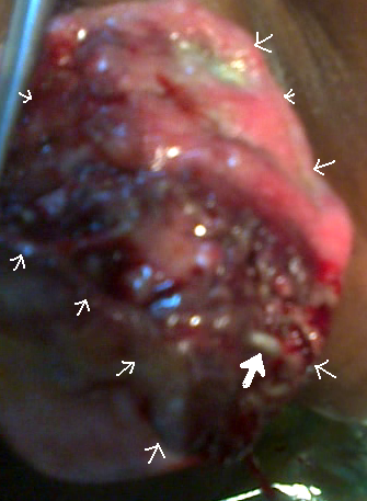
Free-Floating Intraperitoneal Mass
Gupta P, Parulekar SV.
Genital Myiasis
Dhakne P, Gupta AS.
Intramyometrial pregnancy
Kumari M, Gupta AS.
Forgotten Vaginal Ring Pessary
Khadkikar R, Panchbudhe S, Channawar S, Chauhan AR.
Difficult Vaginal Hysterectomy: A New Approach
Parulekar SV
Editorial
Parulekar SV
Ovarian Failure with Uterine Artery Embolization
Mirchandani A, Parulekar SV.
Hysterographic Assessment of Repair of Uterine Chronic Rupture
Sharma G, Samant PY, Parulekar SV.
Ectopia Vesicae and Intrauterine Pregnancy
Mahajan J, Samant PY, Parulekar SV.
Free-Floating Intraperitoneal Mass
Gupta P, Parulekar SV.
Genital Myiasis
Dhakne P, Gupta AS.
Intramyometrial pregnancy
Kumari M, Gupta AS.
Forgotten Vaginal Ring Pessary
Khadkikar R, Panchbudhe S, Channawar S, Chauhan AR.
Difficult Vaginal Hysterectomy: A New Approach
Parulekar SV
Editorial
Parulekar SV
Editor-in-Chief
Editor-in-Chief
In a busy hospital practice, one gets to see a lot of
patients. When the hospital is a tertiary level care center, like our
center,
one encounters a number of unusual cases. There is a lot to be learned
from
such cases, because their presentation and management are different
from the
descriptions in the standard textbooks. Unless these cases are
documented, they
tend to be forgotten, until one encounters a similar case again.
Without
documentation, knowledge of these cases is not available to others in
the
scientific community. Case reports need to be published so that these
two
deficiencies in learning are overcome. Then one day someone can review
the
literature and compile a series of all such cases, so that
statistically
significant conclusions can be drawn. Science makes progress in this
way.
There is another deficiency in
learning. Practice of
conventional Gynecology and Obstetrics is based on textbook teaching
and
management algorithms. But new ideas are required for science to
progress. Many
people get new ideas. These ideas can be on anything – clinical tests,
laboratory tests, instrumentation, or operative procedures. A few of
these are
brilliant. These thinkers need a platform to present their ideas, even
before
they go through the process of clinical studies, so that there can be
brainstorming on them. The final version can then be put to critical
evaluation
in a scientific study.
Journal
of Postgraduate Gynecology & Obstetrics is launched with these
objectives
in mind. We believe in free education for all, including publication of
scientific content. Hence we have made this journal open source. Unlike
many
such journals on the net, it is totally free. There are no hidden
charges, like
processing fee, printing fee, color illustration charges etc. It is
perhaps the
only journal that readers can access for free and contributors can
contribute
to for free. This should help all those in the less privileged
countries,
though people in the privileged countries are also welcome to these
resources.