Volume 4 Issue 11, November 2017

Editorial

Gupta AS

Medical disorders in pregnancy is a reality for the practicing obstetrician. Most of the times medical disorders in a gravida are diagnosed in the antenatal period as more and more women are registering and following up in the antenatal period. However, in a significant number these disorders are still diagnosed in labor or in the postpartum period. In such emergency situation the obstetrician has to depend on self to confirm and or manage these conditions which may have adversely or positively affected pregnancy or labor. Obtaining a physician or a cardiologists consultation may not always be feasible in emergency hours.

Cardiac disorders in pregnancy is one such medical disorder frequently suspected or diagnosed by the obstetrician as many women visit a doctor for the first time during their pregnancy due to the social structure of the Indian society. Incidence of cardiovascular disorders in pregnancy ranges from 1 to 4 %. Mortality attributed to cardiac disorders is about 10 %. Even though in the western world congenital heart diseases form the bulk in India rheumatic valvular lesions still predominate. Another category of ischemic heart disease that was previously not seen in Indian pregnant population is now seen in the pregnant women, probably due to late marriages, late child birth, obesity, diabetes mellitus either preexisting or gestational and hypertension. All these patients require multidisciplinary management

The cardiologist and the obstetricians also face the task to identify those cardiac cases who should not conceive or should conceive after cardiac surgery or stabilization of their cardiac condition. Some of the cardiac conditions wherein pregnancy is contraindicated includes primary pulmonary hypertension, critical mitral stenosis or aortic stenosis, certain cases of cardiomyopathy wherein the ejection fraction is less than 30 %, uncorrected coarctation of the aorta especially if associated with aneurysm. Since many times the obstetrician sees the patient first only in pregnancy the cardiologist and the physicians have a main role to play in imparting contraceptive counseling or referring them to a contraceptive provider and convince the couple to either avoid pregnancy totally or to defer it at least till a definitive correction of her cardiac lesion has been done. Unfortunately this is not the norm and the social fabric of our society pressurizes this woman to conceive in poor general health.
When such a woman conceives; the obstetrician is faced with the daunting task of managing a very high risk pregnancy with significant chance of maternal and perinatal morbidity or mortality. Sometimes if these women present in the first trimester of pregnancy, termination of pregnancy can be done. Both these scenarios can be avoided if medical providers of all specialties and sub specialties realize their responsibility and counsel the couple with contraceptive advises during their reproductive life rather then leaving this task to the obstetrician, gynecologist or family physicians.  

This November issue of the journal has three cardiac cases with pregnancy. Pregnancy should have been avoided in one woman who had to undergo mitral valve replacement as an emergency procedure in pregnancy and the remaining two were diagnosed in their current pregnancy with life threatening cardiac condition. Fortunately all three women survived their condition and were counseled to avoid pregnancy in future or to consider it only after confirming with the cardiologist.

Blanching Test To Locate An Intramural Leiomyoma

Innovation
Author Information

Parulekar SV
(Professor and Head, Department of Obstetrics and Gynecology, Seth G S Medical College & KEM Hospital, Mumbai, India.)

Abstract

Location of a leiomyoma may be clinically evident when it is submucous or subserous pedunculated. It is more difficult to locate it clinically as well as during a myomectomy operation when it is intramural. A new technique is described here to help locate the leiomyoma when the uterus is enlarged uniformly and symmetrically.

Introduction

An intramural leiomyoma enlarges the uterus. It may be an asymmetrical enlargement if it is on one side, anterior or posterior, right or left.[1,2] Usually it can be located easily during a myomectomy operation when it alters the contour of the uterus asymmetrically, and is firm in consistency. However it may not be possible to locate it if it is deep seated and causes uniform and symmetrical enlargement of the uterus. It is even more difficult to locate it if it is soft in consistency. A new method to help locate such a leiomyoma during myomectomy.

Method

The part of the uterus suspected to have the intramural leiomyoma is held between two fingers of each hand on either side, making pressure towards each other and also below the leiomyoma, so that it tends to get displaced towards the surface of the uterus. The color of the uterine surface is observed prior to making the pressure, during maintenance of the pressure, and after relieving the pressure. If there is no blanching of the surface at any time, there is no leiomyoma underneath the uterine surface at that site. If there is blanching with pressure, and restoration of original color after the pressure is removed, there is an intramural leiomyoma deep to the surface of the uterus under study.


Figure 1. Intraoperative view of the uterus. The uterus seems to be uniformly and bilaterally symmetrically enlarged. There is no blanching of any part of the uterine serosa.


Figure 2. Pressure on the posterior wall of the corpus to demonstrate a leiomyoma on the right side, if any. There is no blanching of uterine serosa.


Figure 3. Pressure on the posterior wall of the corpus to demonstrate a leiomyoma on the left side, if any. There is blanching of uterine serosa.


Figure 4. Presence of a leiomyoma at the site suggested by the blanching test is confirmed by extrusion of the leiomyoma through an incision in the uterine wall and pseudocapsule of the leioyoma.

Discussion

Usually a leiomyoma is of a consistency different from that of the myometrium. It is a rounded structure. It is firmer and can be palpated through the myometrium lying over it.[1,2] When it lies deeper and is softer due to degenerative changes, it does not offer greater resistance to palpation than the overlying myometrium.[3] A softer consistency may be due to degeneration, cellular nature of a leiomyoma, or a sarcoma. A leiomyoma is best located by an ultrasonography preoperatively. When in doubt, a coputed tomography or a magnetic resonance imaging scan help.[4-8] But even with that knowledge, it is important to locate it intraoperatively so that the incision on the uterus can be made at a site best suited for removal of the leiomyoma.[9,10] An incision placed away from such a site will involve having to make bigger incisions, greater degree of cutting into the myometrium, greater amount of intraoperative blood loss and use of more suture material for closing the incision.

The method described here depends on blanching of the overlying myometrium by the pressure of an underlying leiomyoma. When the leiomyoma is made prominent by compressing the myometrium on its sides, it rises towards the surface of the uterus and presses on the myometrium lying over it. Since it is already less vascular due to the pressure of the underlying leiomyoma before the test is performed, the surface does not blanch. But when the test is done in an area of the uterus that does not have an underlying leiomyoma, the surface of the uterus blanches and goes back to the original color when the pressure on the leiomyoma from two sides is removed.

This is a simple, noninvasive test that can be performed during both a laparotomy and laparoscopy. In case of the former, fingers of two hands are used, while in case of the latter terminal portions of  two grasping forceps are used. This test should prove a useful adjunct to conventional methods like palpating directly over the leiomyoma and observing the contour of the uterus for distortion.

Acknowledgment

I thank Dr Durga Valvi for taking the intraoperative photographs.

References
  1. Novak ER, Woodruff JD. In Gynecologic and Obstetric Pathology. 8th ed. Philadelphia, USA: W.B. Saunders; 1979. Myoma and other benign tumors of the uterus; pp. 260–78.
  2. Prayson RA, Hart WR. Pathologic considerations of uterine smooth muscle tumors. Obstet Gynecol Clin North Am. 1995;22:637–57.
  3. Buttram VC, Jr, Reiter RC. Uterine leiomyomata: Etiology, symptomatology, and management. Fertil Steril. 1981;36:433–45.
  4. Stephen Karasick, Anna S Lev-Toaff, Michael E Toaff. Imaging of uterine leiomyomas: Pictorial essay. Am J Roentgenol. 1992;158:799–805.
  5. Vitiello D, McCarthy S. Diagnostic imaging of myomas. Obstet Gynecol Clin N Am. 2006;33:85–95.
  6. Ascher SM, Silverman PM. Applications of computed tomography in gynecologic diseases. Urol Radiol. 1991;13:16–28.
  7. Stamatopoulos CP, Mikos T, Grimbizis GF, et al. Value of magnetic resonance imaging in diagnosis of adenomyosis and myomas of the uterus. J Minim Invasive Gynecol. 2012;19(5):620–626.
  8. Borgfeldt C, Andolf E. Transvaginal ultrasonographic findings in the uterus and the endometrium: Low prevalence of leiomyoma in a random sample of women age 25–40 years. Acta Obstet Gynecol Scand. 2000;79:202–7.
  9. Hurst BS, Matthews ML, Marshburn PB. Laparoscopic myomectomy for symptomatic uterine myomas. Fertil Steril. 2005;83:1–23.
  10. Manyonda I, Sinthamoney E, Belli AM. Controversies and challenges in the modern management of uterine fibroids. BJOG. 2004;111:95–102.
  11. Daniell JF, Gurley LD. Laparoscopic treatment of clinically significant symptomatic uterine fibroids. J Gynecol Surg. 1991;7:37–9.
Citation

Parulekar SV. Blanching Test To Locate An Intramural Leiomyoma. JPGO 2017. Volume 4 No. 11. Available from: http://www.jpgo.org/2017/11/unusual-broad-ligament-leiomyoma.html

Unusual Broad Ligament Leiomyoma

Author Information

Swaminathan G*, Parulekar SV**.
(* Specialty Medical Officer, ** Professor and Head, Department of Obstetrics and Gynecology, Seth G S Medical College & KEM Hospital, Mumbai, India.)

Abstract
Extrauterine leiomyomas may develop in the broad ligament or at any other site where smooth muscle is present. Primary broad ligament tumors are rare and these pose diagnostic difficulties due to their rarity. Broad ligament is the most common extrauterine site for the occurrence of a leiomyoma. We report a case of a 47 year old woman with a large right sided true broad ligament leiomyoma. On the basis of clinical examination and ultrasound, a provisional diagnosis of uterine leiomyoma was made. On laporotomy, it was found to be a large 18*15*10 cm size leiomyoma in the right broad ligament for which enucleation of leiomyoma was done followed by total abdominal hysterectomy.

Introduction

Leiomyomas are the most common uterine tumors, most commonly found during the reproductive years. They are smooth muscle tumors which are benign in nature. They can be either  intrauterine or extrauterine. The classification is mainly determined by the site of its origin and the direction of growth. They are broadly classified into three main types :  interstitial, subserous and submucous. Entities called as extra-uterine leiomyomas exist but they are not as common as uterine leiomyomas. Extra-uterine leiomyomas may develop in the broad ligament or at any other site where smooth muscle is present.[1] Primary broad ligament tumors are rare. They pose specific diagnostic difficulties due to their rarity. Tumors of broad ligament are mostly epithelial in nature and mesenchymal tumors are less common. The commonest mesenchymal tumors found in the broad ligament are leiomyomas.[2] The true incidence of broad ligament leiomyomas is not known. They can cause a variety of symptoms like menstrual irregularities and pressure effects.[1,2] Usually the diagnosis of broad ligament leiomyomas can be made clinically and confirmed by imaging. If not, it is evident during a laparotomy. We present a case in which it was missed clinically, by imaging and was very difficult during a laparotomy.

Case Report

 A 47 year old para 3 living 3 with previous 3 full term normal deliveries came to our outpatient clinic with complaints of abnormal uterine bleeding. She had complaints of menorrhagia since 3 cycles with menses every 15-20 days lasting for 5-6 days with soakage of 5-6 pads per day and passage of clots. She also had complaints of pain in abdomen in right flank and hypogastric region which was dull aching in nature since 15-20 days. She also had frequency of micturition for 1 month. On examination her general condition was fair, vital parameters were within normal limits, and there was no abnormality on systemic examination. On abdominal examination, there was a nontender lump of around 16-18 weeks of gestation arising from the pelvis. On per speculum examination, cervix and vagina were healthy and there was minimal bleeding through os. On bimanual pelvic examination, a single large intramural leiomyoma 5-6 cm in diameter was felt posterolaterally on right side. Ultrasound scan of pelvis was suggestive of an enlarged uterus with multiple intramural uterine leiomyomas. A provisional diagnosis of uterine leiomyomas with abnormal uterine bleeding was made and decision was taken for total abdominal hysterectomy with bilateral salpingectomy. Investigations for fitness for anesthesia were normal. A laparotomy was done. Intraoperatively, there was a large 10x12 cm sized mass on the right side and midline. The uterus and adnexal structures were not found. We were called for assistance at that stage. We found that the mass was covered by peritoneum and had large vessels on the surface. It was not mobile, which suggested that it was extraperitoneal. Its relationship to the uterus was not apparent because the uterus was not seen. Further examination after enlargement of the abdominal incision showed that the uterus was of normal size and shape and was situated within the pelvis, below and to the left of the mass. There was no apparent connection between the uterus and the mass. The fallopian tubes and ovaries were normal. A diagnosis of a right sided true broad ligament leiomyoma was made. 


Figure 1. Showing uterus (U) and right broad ligament leiomyoma (M), ovaries (O) and fallopian tubes (F), which have been dragged up by traction with a clamp applied to the left cornual structures.

The right broad ligament was opened by clamping, cutting and ligating the right round ligament. The leiomyoma was dissected and removed after locating the right ureter, which was found to be below and lateral to the leiomyoma. Then total abdominal hysterectomy and bilateral salpingectomy was done. Hemostasis was achieved in the bed of the leiomyoma. A drain was kept in the right broad ligament and the abdomen was closed. The patient made an uneventful recovery. The leiomyoma weighed 1150 g. Histopathological examination confirmed the diagnosis of a uterine leiomyoma. The uterus and fallopian tubes showed normal histology.


Figure 2. Enucleation of broad ligament leiomyomyoma.


Figure 3. Broad ligament leiomyoma specimen.

Discussion

Broad ligament is a peritoneal fold  consisting of two layers that connects the sides of uterus to the lateral walls of the pelvis and its floor.[3] Epithelial tumors are the commonest broad ligament tumors, whereas mesenchymal tumors are extremely rare. Among the mesenchymal tumors, the commonest one is leiomyoma.[4] Broad ligament leiomyomas are of two types. A primary/true broad ligament leiomyoma arises from the smooth muscle fibers that are normally found in the tissues in the broad ligament. It can attain a very big size and may distort the fallopian tube and compress the ureter causing hydroureter and hydronephrosis. But it is entirely separate from the uterus. Hence it can displace the uterus, but not distort it. The anatomical relationship of the uterine vessels and ureter to such a leiomyoma is variable. Hence there is serious risk of injury to the ureter if it is not located and protected prior to clamping, cutting or ligating anything in the broad ligament. A secondary/false broad ligament leiomyoma arises mostly from the lateral walls of the uterus or the cervix and grow laterally between the two layers of the broad ligament but retains its attachment to the uterus. The uterine vessels and ureter lie lateral to the tumor.[5] Such a tumor has a pseudocapsule. Hence when it is enucleated within the pseudocapsule, there is no risk of injury to the ureter.
Making a diagnosis of broad ligament leiomyomas is always a challenge.[6] The most useful modalities for diagnosis are ultrasound (USG), computed tomography (CT) and magnetic resonance imaging (MRI). On imaging, “bridging vessel sign” is helpful in the diagnosis of a leiomyoma.[2] Transvaginal ultrasound can diagnose broad ligament leiomyoma because it allows clear visual separation of the uterus and ovaries from the mass. On ultrasound, a broad ligament leiomyoma usually presents as a well circumscribed hypoechoic solid mass with a whorled appearance, with variable echogenicity depending on the extent of calcification and degeneration. Pedunculated leiomyomas may be mistaken for solid ovarian masses on ultrasound, MRI is indicated in such circumstances.[7] On MRI, broad ligament leiomyomas have the typical appearance of leiomyomas and are seen as sharply emarginated lesions of low signal intensity on both T1W and T2W sequences due to their high fibrotic content. They are usually not surrounded by a pseudocapsule and do not show interface vessels which are imaged as perilesional rim enhancement. The differential diagnosis is with parasitic leiomyoma of the broad ligament, subserosal leiomyomas, masses of ovarian origin, fibrotic adnexal lesions and other solid tumors from the broad ligament. Parasitic leiomyomas of the broad ligament usually originate from the uterus and they invade the broad ligament, additionally recruiting own arterial supply, and either maintaining or losing their original uterine attachment. Pedunculated subserosal leiomyomas, may be hard to distinguish from broad ligament leiomyomas; thus identification of the site of attachment is crucial to establish the uterine origin. Differentiation of a true broad ligament leiomyoma from other pelvic extraperitoneal tumors is essential for the patients’ further management.

It was a large true broad ligament leiomyoma in our case. Broad ligament leiomyomas have the potential to grow to a large size[8]. We did not do CT or MRI because the diagnosis was apparently clear clinically and was confirmed by USG. Routine use of CT and MRI is not possible in resource poor healthcare facilities. The nature of the tumor could not be determined by the operating surgeon because the uterus and adnexal structures were not seen anywhere. Finally it was found that the leiomyoma was growing from above the level of the uterine vessels and had continued to grow cranially after displacing the normal-sized uterus and its adnexal structures downward and to the left. These structures were hidden below the large mass. Since they serve as a landmark for a gynecologist while dealing with intraperitoneal as well as extraperitoneal pelvi-abdominal masses, their localization is an important step in the management of such masses. Surgery is challenging in case of broad ligament leiomyomas because of their location and size, especially since surrounding organs like ureters, urinary bladder and intestines may be at risk. It is very important to identify the ureteric course during surgery. Also, leiomyoma may be adherent to surrounding structures and therefore lead to greater risk of inadvertent injury.[1]

Conclusion

True broad ligament leiomyoma is a tumor that can be challenging  in both its diagnosis and management. Identification of the uterus and adnexal structures is crucial in determining the source of the tumor during a laparotomy. Though imaging modalities are helpful, the final diagnosis needs to made intraoperatively and confirmed by histopathological examination.

References
  1. Fasih N, Prasad Shanbhogue AK, Macdonald DB, Fraser-Hill MA, Papadatos D, Kielar AZ, et al. Leiomyomas beyond the uterus: unusual locations, rare manifestations. Radiographics. 2008;28:1931–1948.
  2. Bansal P, Garg D. A case of massive broad ligament leiomyoma imitating an ovarian tumour. J Clin Diagn Res 2014;8:136-7.
  3. Buttram VC Jr, Reiter RC. Uterine leiomyomata: etiology, symptomatology, and management. Fertil Steril 1981;36:433.
  4. Thor AD, Young RH, Clement PB. Pathology of fallopian tube, broad ligament, peritoneum and pelvic soft tissues. Hum Pathol 1991;22: 856–67.
  5. Pandit P, Chandak S. Laparoscopic Management of Broad Ligament Fibroids. Journal of Gynecological Endoscopy and Surgery. 2011;2(1):64-66.
  6. Stewart EA. Uterine Fibroids. The Lancet, 2001;357, 293-298.
  7. Fasih N, Prasad Shanbhogue AK, Macdonald DB, Fraser-Hill MA, Papadatos D and Kielar AZ. Leiomyomas beyond the Uterus: Unusual Locations, Rare Manifestations. Radiographics, 2008;28, 1931-1948.
Citation

Swaminathan G, Parulekar SV. Unusual Broad Ligament Leiomyoma. JPGO 2017. Volume 4 No. 11. Available from: http://www.jpgo.org/2017/11/unusual-broad-ligament-leiomyoma.html

Successful Maternal Outcome In Pregnancy With Pulmonary Hypertension

Author Information

Madhva Prasad S,  Gupta AS**
(* Assistant Professor, ** Professor. Department of Obstetrics and Gynecology, Seth GS Medical College and KEM Hospital, Mumbai, India.)

Abstract

A 20 year old G2 A1 presented at 27 weeks with sudden breathlessness and was found to have severe pulmonary hypertension. She was managed with diuretics and required sildenafil.  She was anti-β2 glycoprotein 1 positive. She went into spontaneous preterm labor. Maternal outcome was good. A successfully managed parturient with severe pulmonary hypertension and related issues are discussed here.

Introduction

Pulmonary hypertension as a consequence of preexisting cardiac valvular disorders is known as Eisenmenger syndrome. However, when it occurs with no other coexisting cardiac lesions, it is called primary pulmonary hypertension. It is a disorder which portends poor maternal and neonatal outcomes.

Case Report

A 20 year old woman married for 2 years gravida 2 abortion 1 with 27 weeks gestation presented to the emergency with complaints of worsening breathlessness over 2 days and occasional pain in the abdomen. She was apparently asymptomatic till 2 days prior and currently had dyspnea on exertion (NYHA grade III). She did not have any swelling, decreased urine output, palpitations, faintness, cough or fever. She was registered with a private practitioner and had 2 antenatal visits. Ultrasonography at 18 weeks showed no malformations but oligohydramnios, for which no specific therapy had been initiated. On arrival, she was evaluated and found to be uncomfortable at rest. She appeared to have peripheral cyanosis. Pulse was 98 beats per minute, blood pressure was 100/60 mm Hg and on auscultation S1 and S2 were well heard, and no murmurs were audible. Respiratory rate was 24 per minute and oxygen saturation on finger probe was 83%. Abdominal examination showed relaxed uterus of 26 weeks gestation with audible fetal heart sounds. On per vaginal examination, internal os was closed and cervical length was adequate. ABG revealed oxygen saturation of 91% (on oxygen).  Immediately, nasal oxygen and diuretics (tablet furosemide 40 mg) were started. Cardiologist evaluated her and did her echocardiography. It showed a pulmonary artery pressure of 120 mm Hg, suggestive of pulmonary hypertension and ejection fraction of 50 %. It also showed, “D” shaped left ventricle, right atrial and right ventricular volume overload and moderate pulmonary regurgitation. Diuretics were continued and antibiotics were started. Cardiologist started tablet sildenafil 10 mg twice a day for pulmonary hypertension. Iron and calcium were continued. Over the next few days, her respiratory status improved and she was slowly weaned off oxygen. Upon stabilization, work up for associated conditions was done. 
Hemoglobin was 12.9 gm%, platelet count was normal. Liver and renal function tests were normal. TSH was 2.85 U/ml. Autoimmune workup showed anti-β2 glycoprotein 1 IgM positive, RA factor was negative, C3, C4 were normal, ANA, ANCA, LA were all negative. In view of antibody positivity, tablet aspirin 75 mg was started. Ultrasonography showed fetal intrauterine growth restriction with doppler flows suggestive of a raised S/D ratio and intermittent loss of diastolic flow. 
She was kept under observation in the ward. She went into spontaneous preterm labor at 28 weeks and progressed spontaneously. Since steroids were already completed, no tocolysis was given. Strict monitoring of saturation, nasal oxygen, 2 hourly ABG monitoring and fluid restriction was followed. Cardiologist reassessed the patient and advised to allow vaginal trial due to advanced labor with imminent delivery.  She delivered in four hours vaginally a male child of 956 grams with Apgar scores of 7/10 and 9/10. Baby required 15 days' NICU admission but no invasive ventilation. 
Mother was transferred back to ICU for observation, and she did not have any worsening of symptoms. Sildenafil was continued. Repeat echocardiography showed pulmonary artery pressure of 85 mm Hg. She was discharged on day 15 of delivery with advice to follow up regularly. She chose barrier contraception. 

Discussion

Pregnancy with pulmonary arterial hypertension (PAH) are associated with multiple maternal and neonatal risks. It is classified by the WHO risk classification as “very high risk for maternal mortality or severe morbidity wherein pregnancy is contraindicated and termination should be discussed”.
Pulmonary arterial hypertension is defined as a mean pulmonary pressure of above 25 mm Hg. Class 1 or primary pulmonary hypertension is used to described those where no specific cause is found, or is associated with some connective tissue disorder. When pulmonary artery pressure is > 50 mm Hg, it is termed as severe.  Our patient had pulmonary artery pressure of 120 mm of Hg, and had associated anti-β2 glycoprotein 1 IgM positive status. Hence our patient was designated as severe primary pulmonary hypertension.[1, 2] 
Very few studies have documented the outcomes among pregnancies with PAH. In a study by Duarte et al, those who were diagnosed early and underwent pregnancy termination at around 13 weeks had good maternal outcome. However, our patient first presented to us only at 27 weeks of gestation. Scant data exist regarding how many patients in the developing world utilize medical services of early termination in such scenarios.[3] Even when termination is offered, patients may exercise autonomy and refuse the same; as described by Terek et al.[4]
If patients present preconceptionally and insist on pregnancy, regular prenatal and antenatal care is a must.[5] In the study by Duarte et al also, patients who were continuing pregnancy were hospitalized around 27- 30 weeks. Ultrasonography showed IUGR and doppler showed absent end-diastolic flow in our patient; however, she was put on surveillance with weekly doppler for fetal indication. A higher occurrence of SGA babies has been noted in the study by Subbaiah et al.[2] 
The medical management of primary pulmonary hypertension consists of sildenafil, intravenous prostanoids, combination therapy and newer molecules like bosentan. Though it is not yet widely approved for use in pregnancy, recent meta analyses are slowly establishing that the drug is well tolerated and appears to be safe in pregnancy; with no increase in adverse maternal or perinatal outcomes.[6]
While intravenous prostanoids is an option in the management of pulmonary hypertension, pregnancy precludes its use due to its effects on the uterus. Our patient had a spontaneous precipitate preterm labor, and her condition did not worsen. As in the study by Duarte et al, all patients were managed by cesarean sections, which is supported by the fact that it avoids prolonged labor and unpredictable maternal hemodynamics. However, there is a raised risk of death following cesarean section. Hemnes et al have provided a review of the available literature on pregnancy with pulmonary hypertension. Though no universally accepted guidelines exist regarding mode of delivery, appropriate individualized decisions based on sound clinical judgments must be done till further data is available.[7,8,9] 
To conclude, multidisciplinary management of patients with involvement of obstetrician and cardiologists, successful management of patients with pulmonary hypertension can be done. Sildenafil appears to be well tolerated. 

References 
  1. Cardiovascular disorders. In Cunningham FG, Leveno KJ, Bloom SL, Spong CY, Dashe JS, Hoffman BS, editors. Williams’ Obstetrics. 24th ed.  New York: Mc Graw hill 2014;   Pg 977. 
  2. Subbaiah M, Kumar S, Roy KK, Sharma JB, Singh N. Pregnancy outcome in women with pulmonary arterial hypertension: single-center experience from India. Arch Gynecol Obstet. 2013;288(2):305–9.
  3. Duarte AG, Thomas S, Safdar Z, Torres F, Pacheco LD, Feldman J, et al. Management of pulmonary arterial hypertension during pregnancy: a retrospective, multicenter experience. Chest. 2013;143(5):1330–6.
  4. Terek D, Kayikcioglu M, Kultursay H, Ergenoglu M, Yalaz M, Musayev O, et al. Pulmonary arterial hypertension and pregnancy. J Res Med Sci. 2013;18(1):73–6. 
  5. Bao Z, Zhang J, Yang D, Xu X. [Analysis of high risk factors for patient death and its clinical characteristics on pregnancy associated with pulmonary arterial hypertension]. Zhonghua Fu Chan Ke Za Zhi. 2014;49(7):495–500. Available from: http://www.ncbi.nlm.nih.gov/pubmed/25327730
  6. Dunn L, Greer R, Flenady V, Kumar S. Sildenafil in Pregnancy: A Systematic Review of Maternal Tolerance and Obstetric and Perinatal Outcomes. Fetal Diagn Ther. 2017;41(2):81–8. 
  7. Hemnes AR, Kiely DG, Cockrill BA, Safdar Z, Wilson VJ, Al Hazmi M, et al. Statement on pregnancy in pulmonary hypertension from the Pulmonary Vascular Research Institute. Pulm Circ. 2015; 5(3):435–65. 
  8. Bédard E, Dimopoulos K, Gatzoulis MA. Has there been any progress made on pregnancy outcomes among women with pulmonary arterial hypertension? Eur Heart J. 2009;30(3):256-65. 
  9. Khan J, Idrees MM. Saudi Guidelines on the Diagnosis and Treatment of Pulmonary Hypertension: Pregnancy in pulmonary hypertension. Ann Thorac Med. 2014 Jul;9(Suppl 1):S108-12.
Citation

Madhva Prasad S, Gupta AS. Successful Maternal Outcome In Pregnancy With Pulmonary Hypertension.  JPGO 2017. Volume 4 No.11. Available from: http://www.jpgo.org/2017/11/successful-maternal-outcome-in.html

Successful Outcome With Mitral Valve Replacement In Pregnancy

Author Information

Harsha A*,  Pardeshi S**, Gupta AS***.
(* Second Year Resident, ** Assistant Professor, *** Professor. Department of Obstetrics and Gynecology, Seth GS Medical College and KEM Hospital, Mumbai, India.)

Abstract

Rheumatic mitral stenosis constitutes a major cause of acquired heart disease complicating pregnancy in India. Here, a rare case of a pregnant woman undergoing balloon mitral valvuloplasty, for severe mitral stenosis, leading to rupture of mitral valve leaflet and eventually surviving mitral valve replacement is being discussed, she could continue the pregnancy till term and had an elective cesarean section.

Introduction 

Heart disease complicates nearly 1% of all pregnancies and now is one of the leading causes of indirect maternal mortality.[1]  In presence of maternal heart disease, the circulatory changes may lead to death of the mother or the fetus, cardiac failure being most common cause of maternal mortality in such cases. Rheumatic heart disease is the most common heart disease in pregnancy and otherwise in developing counties.[2]

Case Report  

A 27 years old female, married since 9 years, with previous lower segment cesarean section (for meconium stained amniotic fluid), at 25 weeks and 3 days of gestation with critical mitral valve stenosis came to emergency with complaints of  grade 4 breathlessness. On examination, pulse rate was 100 per minute, blood pressure was recorded to be 90/70 mm of Hg. In cardiovascular system examination, first and second heart sounds were heard normally along with a mid diastolic murmur and pansystolic murmur. All lung fields were clear and air entry was bilaterally equal on respiratory system examination. She was a diagnosed case of rheumatic heart disease, having severe mitral valve stenosis (MS), with severe mitral valve regurgitation (MR), moderate tricuspid valve regurgitation (TR), and severe pulmonary hypertension (PH), diagnosed during her previous pregnancy, 8 years back. She was admitted, 2-D echocardiography was done. The ejection fraction was 60%, mitral valve area was 0.7/0.8 mm2 , it showed severe MR (central jet), severe MS, severe TR and severe PH. She was stabilized and started on antidiuretics. She underwent balloon mitral valvuloplasty, during which she sustained left commissural and anterior leaflet rupture, she became unstable, and an emergency mitral valve replacement (MVR) had to be done for her. Native mitral valve was excised, MVR was done using Sorin 25 mm mechanical valve following which she was kept under observation in cardiac ICU for 5 days on ventilator support. She was put on digoxin, frusemide, spironolactone and aspirin. For anticoagulation, she was put on warfarin 5 mg OD and 7.5 mg on two days in a week. She got discharged against medical advice on day 6 of the surgery. She followed up in antenatal OPD fortnightly. In her routine antenatal investigations, fasting blood sugar was 67 mg/dl, and postprandial blood sugar was 104 mg/dl. Her TSH level was 1.52 IU/L. She tested negative for HIV, HBsAg, HCV and VDRL. She received 2 doses of tetanus toxoid. In the malformation scan however, hydrocephalus (isolated) with involvement of the lateral ventricles was identified in the fetus along with a raised amniotic fluid index of 37 cm.
Later she got readmitted at 35 weeks of gestation in preterm labor. Oral warfarin was replaced with intravenous heparin in the dose of 5000 IU qid. APTT was monitored, maintained between 2 to 3 by titrating the dose of heparin to up to 7500 IU qid.
She had polyhydramnios and fetal anomaly scan showed isolated hydrocephalus. Preterm labor did not progress and pregnancy continued till 36 weeks 5 days, when she went into labor. She underwent a LSCS as she was not willing for a trial of vaginal birth after a cesarean section. Heparin was stopped before LSCS and  was restarted after 6 hours post operatively. She gave birth to a 2.308 kg female child who was assessed by the neonatologists. The neonate did not have hydrocephalus and was neurologically normal. She was stable in the post operative period. On day 5 of LSCS, oral warfarin 5 mg OD was restarted, along with overlapping heparin. APTT was monitored. Eventually heparin was omitted and oral warfarin was continued. She was advised to follow up with the cardiologist, neonatologist and us. Sutures were removal on day 14. Wound had healed well. 

Discussion

Pregnancy induced physiological changes in cardiovascular system has a profound effect on underlying heart disease. Cardiac output increases by more than 40%, heart rate increases and there is a decrease in both systemic and pulmonary vascular resistance.[2] Almost half of the increase in the cardiac output takes place by as early as 8 weeks and all of it by mid pregnancy. Increase in the cardiac output continues to occur throughout pregnancy to up to approximately 40% of the non pregnant state. Women with underlying heart disease are unable to accommodate to these changes and cardiogenic heart failure may occur. Though it is advisable to perform percutaneous balloon mitral valvuloplasty (PBMV) before pulmonary hypertension (PH) has set in, in patients who have MS with established PH, PBMV still remains an effective option with less procedure time.[3] Balloon valvuloplasty may be employed as a bridge to delivery in pregnant women when a definitive corrective surgery in the form of valve replacement is usually done after delivery.[4] There is a substantial risk of fetal death if cardiac operation under cardiopulmonary bypass (CPB) is required, so open heart surgery during pregnancy is suggested only in extreme conditions. Emergency and redo operations have significant surgical risks even under mechanical circulatory support.[5] Second trimester is more appropriate time for cardiac surgery with good fetal prognosis and lesser chance of fetal anomaly, however poor maternal condition may necessitate intervention in early  pregnancy with associated procedural risk to the fetus in the form of anomalies or abortion.[6,7] One such successful case has been published from KEM hospital of mitral valve replacement during pregnancy done at 25 weeks of gestation due to cardiac failure in patient with severe MS with MR with severe pulmonary hypertension, patient survived the procedure, continued pregnancy till term and delivered a live issue by normal vaginal delivery.[8] Fetal prognosis improves if cardiac surgeries are planned later in the pregnancy as reported by Weiss and colleagues.[9] Surgical intervention during pregnancy is done after a serious consideration because of requirement of anticoagulation especially with mechanical prosthetic valves. Porcine valves are preferred during pregnancy.[10] During pregnancy, platelet number and activation of coagulation cascades increases, but contrast fibrinolytic activity decreases. This increases risk of thrombosis in mechanical prosthetic valves (0.7%-6%), so it important to appropriately maintain proper anticoagulation.[11] The procedure of CPB doesn't affect the maternal survival as much as fetal outcome. Maternal outcome in pregnancy is almost equal to that in non pregnant state (1.47%) whereas a fetal mortality of around 16-33% has been still recorded.[12] In a survey done over 35 years in Mayo Clinic 21 pregnant patients who underwent cardiothoracic surgeries during that period were considered.[13] Out of these, 52% of them had premature deliveries and 3 were fetal deaths (14%). The various neonatal complications were growth retardation (5%), respiratory distress syndrome (33%) and developmental delay (14%).[14] There haven't been many studies performed on the fetal circulation during CPB. CPB results in poor placental perfusion and poor respiratory gas exchange due to lower placental flow and pressure along with hypothermia which further worsens the fetal perfusion. [15] Cardiac surgery can be performed with relative safety during pregnancy by adopting normothermic, high flow rate circulation and continuous fetal activity monitoring.[16]

References
  1. Nanna M, Stergiopoulos K.Pregnancy Complicated by Valvular Heart Disease: An Update. J Am Heart Assoc. 2014; 3(3): e000712. 
  2. Nqayana T, Moodley J, Naidoo DP. Cardiac disease in pregnancy. Cardiovasc J Afr. 2008;19(3):145-51.
  3. Ozkan H, Bozat T, Tiryakioglu SK, Ari H. Should we wait until severe pulmonary hypertension develops? Efficacy of percutaneous mitral balloon valvuloplasty in patients with severe pulmonary hypertension: A subgroup analysis of our experience. Cardiol J. 2016;23(2):184-8.
  4. Nwaejike N, Mills K, Stables R, Field M. Balloon aortic valvuloplasty as a bridge to aortic valve surgery for severe aortic stenosis. Interact Cardiovasc Thorac Surg. 2015 Mar;20(3):429-35.
  5. Lin TY, Chiu KM, Shieh JS, Chu SH. Emergency redo mitral valve replacement in a pregnant woman at third trimester: case report and literature review. Circ J. 2008 Oct;72(10):1715-7.
  6. Arnoni RT, Arnoni AS, Bonini RC, de Almeida AF, Neto CA, Dinkhuysen JJ, et al. Risk factors associated with cardiac surgery during pregnancy. Ann Thorac Surg. 2003 Nov;76(5):1605-8.
  7. Becker RM. Intracardiac surgery in pregnant women. Ann Thorac Surg. 1983;36(4):453–8.
  8. Pardeshi S, Mayadeo NM, Warke HS.  Mitral Valve Replacement During Pregnancy. JPGO 2014 Volume 1 Number 4 Available from: http://www.jpgo.org/2014/04/mitral-valve-replacement-during.html
  9. Weiss BM, von Segesser LK, Alon E, Seifert B, Turina MI. Outcome of cardiovascular surgery and pregnancy: a systematic review of the period 1984-1996.Am J Obstet Gynecol. 1998 Dec;179(6 Pt 1):1643-53.
  10. Barbarash L, Rutkovskaya N, Barbarash O, Odarenko Y, Stasev A, Uchasova E. Prosthetic heart valve selection in women of childbearing age with acquired heart disease: a case report.Journal of Medical Case Reports. BioMed Central 2010.2016;10:51.
  11. Roudaut R, Serri K, Lafitte S. Thrombosis of prosthetic heart valves: diagnosis and therapeutic considerations. Heart. 2007;93(1):137–42.
  12. Chambers CE, Clark SL. Cardiac surgery during pregnancy. Clin Obstet Gynecol 1994;37(2):316_23. 
  13. Martin SR, Foley MR. Intensive care in obstetrics: An evidence_based review. Am J Obstet Gynecol 2006;195(3):673_89.
  14. John AS, Gurley F, Schaff HV, Warnes CA, Phillips SD, Arendt KW, et al. Cardiopulmonary bypass during pregnancy. Ann Thorac Surg 2011;91(4):1191-6. 
  15. Assad RS, Lee FY, Bergner K, Hanley FL. Extracorporeal circulation in the isolated in situ lamb placenta: Hemodynamic characteristics. J Appl Physiol 1992;72(6):2176-80. 
  16. Kikon M, Choudhury KD, Prakash N, Gupta A, Grover V, Gupta VK. Mitral Valve Replacement in a Young Pregnant Woman: A Case Report and Review of Literature. Res Cardiovasc Med. 2014; 3(2): e17561.
Citation

Harsha A,  Pardeshi  S, Gupta AS. Successful Outcome With Mitral Valve Replacement In Pregnancy.  JPGO 2017. Volume 4 No.11. Available from: http://www.jpgo.org/2017/11/successful-outcome-with-mitral-valve.html

Fetus Papyraceus In A Twin Pregnancy

Author Information

Daruwale R*, Warke HS. **, Mayadeo NM***.
(* Senior Resident, ** Associate Professor, *** Professor. Department of Obstetrics and Gynecology, Seth GS Medical College and KEM Hospital, Mumbai, India.)

Abstract

When the fetus dies in early pregnancy, the amniotic fluid and placental tissue are absorbed and the fetus is compressed between the membranes with the co-living twin. Such a fetus resembles a parchment paper and is known as fetus papyraceus. We present a case that was diagnosed as dichorionic diamniotic twin gestation in early pregnancy which subsequently resulted in the fetal demise of one twin in second trimester leading to fetus papyraceus and successful healthy outcome of second fetus. 

Introduction 

The term fetus papyraceus is used when intrauterine fetal demise of a twin occurs in early pregnancy, with retention of the fetus for at least 10 weeks resulting in mechanical compression of the small fetus which then resembles a parchment paper.[1] The reported incidence of fetus papyraceus is one in 12,000 pregnancies [2] and approximately 1:190 twin pregnancies.[3] Most of the time, a mummified fetus is an incidental finding during investigation of other pregnancy-related problems on ultrasonography. The complications related to fetus papyraceus depend on chorionicity of the twin pregnancy. Various complications are seen in monochorionic twin pregnancies when compared with dichorionic pregnancies.[3]

Case Report

A 28 year old lady, registered at our institute, gravida 2, para 0, living 0, spontaneous abortion 1 with 38 weeks of gestation came to emergency with complaints of pain in abdomen. Her ultrasonography (USG) done at 8 weeks gestation was suggestive of dichorionic diamniotic twins and all routine blood investigations were within normal limits. Her anomaly scan done at 18 weeks of gestation was suggestive of intrauterine death of one twin and the second twin was alive with no obvious anomalies. Her blood coagulation profile was normal. She and her husband were counseled about her condition and risks to mother and fetus were explained on continuation of the pregnancy. She was managed conservatively with monitoring of coagulation profile every fortnight and ultrasonography every three weeks. Her  sonography at 27 weeks of gestation was suggestive of a single live intrauterine gestation with mummified second twin. She was advised to keep strict count of daily fetal kick movements and report immediately to the emergency room in case of decreased or absent movements. At 32 weeks of gestation she was given two doses of 12 mg betamethasone intramuscular injection 24 hours apart to achieve fetal lung maturity. Weekly non stress tests and biophysical profile were done thereafter to ensure safety of the live fetus.
On examination in the emergency room, her general condition was fair, pulse rate was 88 beats per min, blood pressure was 122/80 mm of Hg. On per abdomen examination, uterus was full term with breech presentation, fetal heart rate was100 beats per minute with variable decelerations but with a good pick up to the baseline. She had two uterine contractions in ten minutes, each lasting for fifteen seconds. On per vaginum examinations, cervix was 2 cm dilated and poorly effaced with breech presentation.
Emergency lower segment cesarean section was conducted in view of fetal distress. A 2.4 kg baby was delivered by breech extraction method.  Baby cried immediately after birth and was shifted to NICU for observation. Intra-operatively, uterus was arcuate  in appearance (figure 1). There was no evidence of septate uterus or vaginal septum. Placenta weighing 900 gm was delivered completely and with membranes. A 6 cm compressed mummified fetus weighing 150 gm was seen along with the membranes on placental examination (figure 2) Rest of the procedure was uneventful. The neonate and the mother were followed for six weeks. There were no complications and she was advised to follow up for one year with the pediatrician to rule out any developmental delay in the baby.


Figure 1. Intraoperative finding of arcuate uterus. 


Figure 2. Fetus papyraceus.

Discussion

Fetus papyraceus is a very rare complication with the incidence of 1 in 12,000 pregnancies. The actual rate of multiple pregnancies is significantly larger than that observed during labor because of the fact that in the course of pregnancy, intrauterine death of one or more fetuses may occur which may be missed.[4] Depending on the gestational period at which fetal death occurs, there are three forms of this complication; vanishing twin syndrome in the first trimester, fetus papyraceus in the second trimester and the macerated twin in the third trimester. In most cases, death occurs in the second trimester. Between 6-8 weeks, the dead embryo in multiple pregnancies may present in the form of a cyst on the fetal surface of the surviving twin’s placenta. After 8 weeks, the death of one embryo, with resorption of amniotic fluid and mummification of the fetal parts, will cause fetus papyraceus.[5] The degree of compression depends on the time span between the fetal death and delivery; the larger the fetus, the more difficult it is to become a fetus papyraceus.[6] Different causes for the intra uterine death of one fetus include twin-twin transfusion syndrome, membranous or velamentous cord insertion, true cord knot, cord stricture, placental insufficiency, and congenital anomalies.[7] 
The risk associated with fetus papyraceus is its effect on mother and surviving co-twin.  Number of the fetuses, gestational age at the time of the death, the reason for the death, the chorionicity and the length of time between demise and delivery of the surviving fetus decides the prognosis of the surviving fetus. In dichorionic twins, the prognosis for the surviving twin is relatively better but in monochorionic twins, the prognosis is poor and is usually associated with neurological damage in the survivor.
Maternal complications associated with fetus papyraceus are preterm labor, infection from a retained fetus, severe puerperal hemorrhage, consumptive coagulopathy, and obstruction by a low-lying mummified twin causing dystocia leading to cesarean delivery.
Close follow up is of utmost importance. Coagulation profile should be checked every 2 weeks and the surviving twin’s well being should be monitored closely with ultrasound, biophysical profile and Doppler. Any anomalies in the surviving twin should be ruled out before making the decision to continue pregnancy. Zygosity and chorionicity evaluation should be performed in the prenatal period. In case of dizygotic twin risk to the surviving twin is not increased and spontaneous onset of labor at term can be awaited under close monitoring of maternal and fetal parameters. In case of monochorionic twin risk to surviving twin is significantly increased through shunt communications which can lead to disseminated intravascular coagulation induced death and organ damage.[8,9]
Neonatal cranial ultrasound is recommended after delivery.[10] Serious compromise in the surviving fetus if anticipated, should be discussed with the parents. Fetal magnetic resonance imaging provides more detailed information about brain lesions in the surviving fetus and its use is recommended.[11] In most of the cases, no complications to the mother or surviving twin are observed and fetus papyraceus is just an incidental finding.  So expectant management with close maternal and fetal surveillance is advised.
Hence timely diagnosis of fetus papyraceus is very important to prevent further complications and have successful surviving fetus outcome. Assuring the parents is very important. Maternal consumptive coagulopathy due to fetal demise of one twin is a rarest reported complication.[12,13]

Conclusion

Though fetus papyraceus is a rare finding, delayed recognition can lead to severe complications. So early diagnosis and documentation is important to prevent the possible complications. Regular antenatal serial ultrasound examinations, serological, hematological, and biochemical testing is required to ensure that the gravida does not develop infection or consumptive coagulopathy. Tests for fetal well-being should also be performed serially. 

References 
  1. Fisk NM. Multiple pregnancies. In Edmonds DK editor. Dewhurst’s Textbook of Obstetrics and Gynaecology for Postgraduates.7th ed, Oxford: Blackwell 1999;298-307.
  2. Bush M, Pernoll ML. Multiple pregnancy. In DeCherney AH, Nathan L editors. Current Obstetric and Gynecologic Diagnosis and Treatment. 10th ed. New York: McGraw-Hill. 2003;315-25.
  3. Leppert PC, Wartel L, Lowman R. Foetus papyraceous causing dystocia: Inability to detect blighted twin antenatally. Obstet Gynecol 1979;54(3):381-83.
  4. Landy HJ, Keith LG. The vanishing twin: A review. Hum Reprod Update 1998;4(2):177-83. 
  5. Daw E. Fetus papyraceus--11 cases. Postgrad Med J. 1983;59(695):598-600. 
  6. Benirschke K. Intrauterine death of a twin: Mechanisms, implications for surviving twin, and placental pathology. Semin Diagn Pathol 1993;10(3):222-31. 
  7. Akbar M, Ikram M, Talib W, Saeed R, Saeed M. Fetus papyraceous: demise of one twin in second trimester with successful outcome of second twin at term. Professional Med J. 2005;12:351-3.
  8. Hagay ZJ, Mazor M, Leiberman JR. Multiple pregnancy complicated by a single intrauterine fetal    death. Obstet Gynecol 1985; 66(6): 837-838.
  9. Hagay ZJ, Mazor M, Leiberman JR, Biale Y. Management and outcome of multiple pregnancies complicated by the antenatal death of one fetus. J Reprod Med 1986; 31(8): 717-720.
  10. Royal College Obstetrician and Gynaecologists. Monochorionic Twin Pregnancy, Management (Green-top Guideline No. 51). November. 2016. https://www.rcog.org.uk/en/guidelines-research-services/guidelines/gtg51/
  11. Righini A, Salmona S, Bianchini E, Zirpoli S, Moschetta M, Kustermann A, et al. Prenatal magnetic resonance imaging evaluation of ischemic brain lesions in the survivors of monochorionic twin pregnancies: report of 3 cases. J Comput Assist Tomogr 2004;;28(1):87-92.
  12. Ong SS, Zamora J, Khan KS, Kilby MD. Prognosis for the co-twin following single-twin death: A systematic review. BJOG 2006;113(9):992-8. 
  13. Novak CM, Patel SV, Baschat AA, Hickey KW, Petersen SM. Maternal coagulopathy after umbilical cord occlusion for twin reversed arterial perfusion sequence. Obstet Gynecol. 2013 Aug;122(2 Pt 2):498-500.
Citation

Daruwale R, Warke HS, Mayadeo NM.  Fetus Papyraceus In A Twin Pregnancy. JPGO 2017. Volume 4 No.11. Available from: http://www.jpgo.org/2017/11/fetus-papyraceus-in-twin-pregnancy.html

Pregnancy Associated Cardiomyopathy

Author Information

Pai K*,  Samant PY**, Khare A***
(* Ex Assistant Professor, ** Professor, *** First Year Resident. Department of Obstetrics and Gynecology, Seth GS Medical College and KEM Hospital, Mumbai, India.)

Abstract

A 21 year old woman presented to emergency medical services on day 3 of spontaneous second trimester abortion and subsequent check curettage. She had complaints of breathlessness, pedal edema and abdominal distension preceding the abortion.  Blood investigations and  echocardiogram  were suggestive of cardiomyopathy due to myocarditis. Cardiac medications including beta blockers, ACE inhibitors and diuretics were started. She symptomatically improved over 1 week.

Introduction

Peripartum cardiomyopathy (PPCM) is a rare dilated cardiomyopathy that causes cardiac failure in women in late pregnancy or early postpartum. Antepartum PPCM is rare. About 90% of the cases are diagnosed in the first two months of the postpartum period. PPCM causes significant morbidity and mortality in both mother and fetus; hence all clinicians and, in particular, emergency physicians should be aware of this disease.  Cardiomyopathy associated with early pregnancy has also been called pregnancy associated cardiomyopathy. 

Case Report

A 21 year old woman who had aborted recently, came to emergency services on day 3 of check curettage with complaints of breathlessness (NYHA grade 2), abdominal distension and bilateral pedal edema since 6 days. She aborted spontaneously at 6 months gestation, which was followed by curettage in a private hospital.  She gave history of blood pressure (BP) reading of 130/90 mm of Hg with no albumen in urine, once during pregnancy for which no medications were given. There was no history of any previous medical or surgical illness. On examination, general condition was stable, vital parameters were normal, there was no murmur on auscultation, bilateral coarse crepitations were present,  jugular venous pressure (JVP) was raised, pedal edema was present. Abdominal examination revealed minimal distension. Uterus was just bulky and non tender, per speculum findings were unremarkable. A 2 D echocardiogram (2D echo) that was done was suggestive of global hypokinesia of the left ventricle, grade 2 diastolic dysfunction with an ejection fraction of less than 30 %.  CPK-MB enzyme level was elevated. Troponin-T was weakly positive. A diagnosis of peripartum cardiomyopathy with probable myocarditis was established on the basis of elevated levels of cardiac markers. She was started on beta blockers, ACE inhibitors and diuretics, inj dobutamine drip, tablet carvedilol and injection furosemide (later converted to tablet furosemide), syrup potassium chloride, tablet spiranolactone, and tablet ramipril. Dosages were titrated as per need. Nebulisation with ipratropium was started. She was also given injection ceftriaxone. She symptomatically improved over one week and a repeat 2D echo showed an ejection fraction of 50 %. Following this, she was discharged on cardiac medications. 

Discussion

PPCM was first described in the 18th century but recognized as a separate clinical entity in 1930 and1971. Demakis et al. described criteria for the diagnosis of PPCM.[1] For diagnosis of PPCM the entity should be seen in the last month of pregnancy or first five months after delivery in patient with no identifiable cause for cardiac failure. There should be no recent history of cardiac disease.  Echo cardiographic finding of severe left ventricular systolic dysfunction, demonstrated by  an ejection fraction of less than 45 % and / or reduced shortening fraction of less than 30 % should be seen. An Indian study showed the incidence of PPCM to be 1 in 1374 live births.[2] PPCM constitutes less then 1 % of all cardiovascular events related to pregnancy.[3] There are multiple risk factors postulated as cause of PPCM which include age over 30 years, multi fetal gestation, multiparity, African descent, maternal cocaine abuse, long term tocolytic therapy and selenium deficiency. Our patient did not have any of the above risk factors. In a series of 123 cases of pregnancy associated cardiomyopathy reported by Elkayam, 23 were before 36 weeks and the earliest was at 17 weeks.[4] Our patient presented around 6 months gestation before pregnancy loss which is very rare.
Though the exact etiology of PPCM is still not known,the following hypotheses are proposed. There is increased susceptibility to viral or autoimmune myocarditis in pregnancy. Raised titres of immunoglobins and other auto antibodies in patients with PPCM are suggestive of abnormal immune response.  Fetal cells enter maternal circulation and remain in circulation without rejection due to weak immunogenic paternal halotype of the chorionic cell. If these cells lodge into the cardiac tissue, it triggers an immune response.[5] Maladaptation to stress of pregnancy; hyperdynamic circulation during pregnancy causing remodeling, thyroid disorder and exaggerated reduction in left ventricular systolic function with stress of gestational hypertension may together contribute to cardiac failure in PPCM. Recent studies implicate the role of a prolactin sub fragment 16-kDa which is cleaved from prolactin due to unbalanced oxidative stress. The 16-kDa can destroy the endothelium and damage the micro circulation in the myocardium, reducing the cardiac function and causing ventricular dilatation.[7] The common symptoms include dyspnea, cough, orthopnea, paroxysmal nocturnal dyspnea and rarely thromboembolic manifestations. The mainstay of investigations includes chest x-ray, electrocardiogram and echocardiography. Cardiac markers, such as Troponin T are  suggested to have prognostic implication. B-type natriuretic peptide (BNP) and N terminal pro – BNP (NT- pro BNP) are recommended by the Heart Failure Association of the ESC Working Group on PPCM.[7] Endomyocardial biopsy may show features of myocarditis, but the decision for biopsy should be taken after discussion with the patient and her family. Viral, bacterial culture, and Coxsackie B titres are done in selected cases. Invasive hemodynamic monitoring shows elevated right and left heart filling pressures with reduced cardiac index.
Management of PPCM is similar to other types of heart failure, apart from concern for the adverse effect of treatment on fetus or breast feeding infant. The aim of therapy in PPCM is to reduce preload, after load and increase the cardiac contractility. These can be achieved by treatment of pulmonary congestion, control of hyper/ hypotension, treatment of cardiac arrhythmias and prevention of thromboembolic events. 
Digoxin and diuretics along with beta-blockers improve left ventricular function in patients of PPCM. ACE inhibitors are the drug of choice in postpartum PPCM. Ventricular arrhythmias should be treated aggressively. Therapy with ionotropes such as dobutamine should be directed by invasive cardiac monitoring. Immunosuppressive therapy may be started after two weeks of standard therapy without response in biopsy proven myocarditis, but still efficacy is unclear. Anticoagulation in PPCM is important as pregnancy itself is a hypercoagulable state, in addition to PPCM with dilatation of heart and turbulent flow of blood. Before delivery, unfractionated or low molecular weight heparin is the choice. In postpartum period warfarin is used.
Occasionally, when medical therapy fails, need for mechanical cardiovascular support (intra-aortic balloon pump, ventricular assisting device) and even cardiac transplant have been reported in the literature.[8]
There is no indication to terminate pregnancy in stable patients. In case of  hemodynamically unstable patients early delivery is recommended.[9,10] Maternal mortality rate is higher in black race, multipara and with LVEF less than 30%. Pregnancy should be avoided if LV ejection fraction is < 25% at diagnosis or with incompletely recovered left heart function.[7] 

Conclusion

Peripartum cardiomyopathy is a rare pregnancy-induced dilated cardiomyopathy, seen in the last month of pregnancy and postpartum period, which makes our case of early onset cardiomyopathy in second trimester a rare one. High index of suspicion is required in women presenting with dyspnea without obvious cause in early pregnancy.

References
  1. Demakis JG, Rahimtoola SH, Sutton GC, Meadws WR, Szanto PB, Tobin JR et al. Natural course of peripartum cardiomyopathy. Circulation. 1971;44(6):1053-61. 
  2. Pandit V, Shetty S, Kumar A, Sagir A. Incidence and outcome of peripartum cardiomyopathy from a tertiary hospital in South India. Trop Doct. 2009;39(3):168-9.
  3. Sliwa K, Damasceno A, Mayosi BM. Epidemiology and etiology of cardiomyopathy in Africa. Circulation. 2005;112(23):3577-834. 
  4. Elkayam U, Akhter MW, Singh H, Khan S, Bitar F, Hameed A, et al. Pregnancy-associated cardiomyopathy: clinical characteristics and a comparison between early and late presentation. Circulation. 2005;111(16):2050-5.
  5. Murali S, Baldisseri MR. Peripartum cardiomyopathy. Crit Care Med. 2005;33(10 Suppl):S340-6.
  6. Hilfiker-Kleiner D, Meyer GP, Schieffer E, Goldmann B, Podewski E, Struman I, et al. Recovery from postpartum cardiomyopathy in 2 patients by blocking prolactin release with bromocriptine. J Am Coll Cardiol. 2007;50(24):2354-5.
  7. Sliwa K, Hilfiker-Kleiner D, Petrie MC, Mebazaa A, Pieske B, Buchmann E, et al. Current state of knowledge on aetiology, diagnosis, management, and therapy of peripartum cardiomyopathy: a position statement from the Heart Failure Association of the European Society of Cardiology Working Group on peripartum cardiomyopathy. Eur J Heart Fail. 2010;12(8):767-78.
  8. Aziz TM, Burgess MI, Acladious NN, Campbell CS, Rahman AN, Yonan N, et al. Heart transplantation for peripartum cardiomyopathy: a report of three cases and literature review. Cardiovasc Surg. 1999;7(5):565-7.
  9. Lee W, Cotton DB. Cardiomyopathy: Current concept and clinical management. Clin Obstet Gynecol. 1989;32(1):54–67. 
  10. Biteker M, Kayataş K, Duman D, Turkmen M, Bozkurt B. Peripartum Cardiomyopathy: Current State of Knowledge, New Developments and Future Directions.Curr Cardiol Rev. 2014; 10(4): 317–326.
Citation

Pai K,  Samant PY, Khare A. Pregnancy Associated Cardiomyopathy. JPGO 2017. Volume 4 No.11. Available from: http://www.jpgo.org/2017/11/pregnancy-associated-cardiomyopathy.html

Anterior Abdominal Wall Reinforcement During Cesarean Section

Author Information

Desai GS*,  Mayadeo NM**.
(* Assistant Professor, ** Professor. Department of Obstetrics and Gynecology, Seth GS Medical College and KEM Hospital, Mumbai, India.)

Abstract

Multiparous women and those with previous cesarean section frequently have a lax abdominal wall with accompanying abdominal wall hernias. Reconstruction is possible during the cesarean section itself and obviates the need for surgery at another time. The authors describe a case in which anterior abdominal wall reinforcement was done for laxity of the anterior rectus sheath of a multiparus woman with a past history of cesarean section. Additionally they discuss on the techniques and methods that can be used to reinforce the anterior rectus sheath. 

Introduction

The last decade has seen the rise in rates of cesarean sections and subsequently the rise in rates of abdominal wall hernia.[1,2]  Repeated surgery also causes weakness of the abdominal wall due to destruction of nerves, muscles and more importantly the rectus sheath. Solution to this is primarily surgical.[3-5] However frequently these women do not visit a surgeon till the time of the next pregnancy where they are scheduled for a repeat cesarean section as in our case. At this point in time a surgeon may or may not be available. Conventional open repair often produces an undesirable scar. Laparoscopic repair requires multiple incisions beyond the umbilicus, specialized equipments, and expensive tissue separating mesh. Correcting the defect at the time of cesarean section avoids need for repeat surgery. The authors write this article with the aim of educating the readers with technique and methods of treating laxity of the abdominal wall at the time of cesarean section. 

Case Report

A 32 year old woman with two living children in her third pregnancy had a satisfactory course during her antenatal checkups. Her first pregnancy resulted in a cesarean section for transverse lie. Apart from this her past history was not significant. Per abdominal examination showed a term pregnancy with a vertical infraumbilical incision. Laxity of the anterior abdominal wall was demonstrable as well as eversion of the umbilicus. She was admitted at 38 completed weeks of gestation for an elective cesarean section with tubal ligation. Written inform consent was taken and patient shifted to the operation theater. After spinal anesthesia the surgical field was disinfected. Vertical skin incision was taken, the incision extended to the limit of the scar of previous pregnancy. Cesarean section was completed and hemostasis achieved. The surgeons decided to tackle the rectus sheath laxity from within. With two Allis' forceps both ends of the rectus sheath were held and rectus sheath was separated from the muscle and subcutaneous tissue. The sheath was then cut and the cut edges sutured over each other in a double breasted manner with monofilament nylon 1-0 continuous interlocking suture material. This was then reinforced with a layer of polyglactin No. 1 continuous interlocking suture. The skin was closed with monofilament nylon 2-0 interrupted suture. At suture removal, two weeks from date of surgery, the she is doing well and the umbilicus is inverted. 


Figure 1. Rectus sheath is exposed after completion of the cesarean section and held with two Allis' clamps.


Figure 2. Rectus sheath is cut into two halves and the Allis' forceps flipped over so that the two halves of rectus sheath overlap each other.


Figure 3. Double breasting of the rectus sheath is done with monofilament nylon 1-0 suture material in a continuous interlocking manner.


Figure 4. Suturing is complete and continued to the entire length of the rectus sheath. 

Discussion

A popular method of treating anterior laxity of the abdominal is by use of a mesh.[6] The mesh can be placed by flipping the anterior abdominal wall over itself with the help of Allis' forceps. The rectus sheath is dissected with the help of either blunt or sharp section and cut longitudinally. The two ends of cut ends of rectus sheath are then approximated with monofilament nylon 1-0 suture. Critical to the approximation is double breasting or overlapping the two leaves of rectus sheath. This tightens the anterior abdominal wall and prevents gaping or giving way of the rectus sheath.
Timing is also of importance. Frequently the surgical correction of the abdominal wall is dissuaded till after the cesarean section. This is not necessary. Correcting the defect at the same time of cesarean section is convenient for both the surgeon and the patient. In our case the skin incision was not extended above the umbilicus and the surgery completed directed from within the peritoneum outwards to the skin.
Laparoscopic repair has today become a popular option for hernia repair. Davis et al compared the laparoscopic and open route. Laparoscopic ventral hernia repair yielded lower rates in terms of postoperative superficial surgical site infections as compared to repair by open surgery.[7] Kitamura et al studied the use of tacks in laparoscopic umbilical hernia repair and found that there are no differences in postoperative complication rates in suture versus tack mesh fixation in laparoscopic umbilical hernia repair.[8] With the advent of natural orifice surgery (NOTES), ventral hernia repair can also be done via the transvaginal route.[9] This however, still remains an uncommon approach for most surgeons in developing countries who primarily rely on a mini laparotomy incision with use of either suturing or a mesh.[10]

Conclusion

Double breasting of the anterior rectus sheath at the time of cesarean section with monofilament non absorbable suture material such as monofilament nylon is required to reinforce the anterior rectus sheath. Alternatives include application of a mesh. 
                                                                                                                                                    References
  1. Betrán AP, Ye J, Moller A-B, Zhang J, Gülmezoglu AM, Torloni MR. The Increasing Trend in Caesarean Section Rates: Global, Regional and National Estimates: 1990-2014. PLoS One. 2016; 11(2): e0148343.
  2. Mittal S, Pardeshi S, Mayadeo N, Mane J.Trends in cesarean delivery: rate and indications. J Obstet Gynaecol India. 2014;64(4):251-254. 
  3. Nockolds CL, Hodde JP, Rooney PS. Abdominal wall reconstruction with components separation and mesh reinforcement in complex hernia repair. BMC Surg. 2014;14(4):25.
  4. Kulaçoğlu H. Current options in umbilical hernia repair in adult patients. Ulus Cerrahi Derg. 2015; 31(3): 157–161.
  5. Korukonda S, Amaranathan A, Ramakrishnaiah VPN. Laparoscopic versus Open Repair of Para-Umbilical Hernia- A Prospective Comparative Study of Short Term Outcomes. J Clin Diagn Res. 2017;11(8):PC22-PC24. 
  6. Cheesborough JE, Dumanian GA. Simultaneous Prosthetic Mesh Abdominal Wall Reconstruction with Abdominoplasty for Ventral Hernia and Severe Rectus Diastasis Repairs. Plast Reconstr Surg. 2015; 135(1): 268–276. 
  7. Davies SW, Turza KC, Sawyer RG, Schirmer BD, Hallowell PT. A comparative analysis between laparoscopic and open ventral hernia repair at a tertiary care center. Am Surg. 2012;78(8):888-92.
  8. Kitamura RK, Choi J, Lynn E, Divino CM.Suture versus tack fixation of mesh in laparoscopic umbilical hernia repair. JSLS 2013;17(4):560-564. 
  9. Jacobsen GR, Thompson K, Spivack A, Fischer L, Wong B, Cullen J, et al. Initial experience with transvaginal incisional hernia repair.Hernia. 2010;14(1):89-91.
  10. Zachariah SK, Kolathur NM, Balakrishnan M, Parakkadath AJ. Minimal incision scar-less open umbilical hernia repair in adults - technical aspects and short-term results. Front Surg. 2014;1:32.
Citation

Desai GS, Mayadeo NM. Anterior Abdominal Wall Reinforcement During Cesarean Section  JPGO 2017. Volume 4 No.11. Available from: http://www.jpgo.org/2017/11/anterior-abdominal-wall-reinforcement.html

Cesarean Scar Endometriosis- A Postoperative Complication On The Rise

Author Information

Kalappa SB*, Tiwari N**, Kolhe A***, Chauhan AR****
(* Third year resident, ** Assistant Professor, **** Professor. Department of Obstetrics and Gynecology, *** Assistant Professor. Department of Pathology, Seth G.S. Medical College and K.E.M. Hospital, Mumbai, India.)

Abstract

Endometriosis is found predominantly in women of the reproductive age group and is defined as the presence of endometrial glands and stroma outside the uterus. The pelvic organs and the peritoneum are the most common sites of implantation. Scar endometriosis is a rare entity caused by the spread of endometrial tissue to the wound at the time of surgery. Here, we present a case of cesarean scar endometriosis in a 36 year old female who had undergone two cesarean sections, the diagnostic approach used and the management options. 

Introduction

The prevalence of endometriosis is assumed to be around 10 % with higher frequency reported in women with pelvic pain or infertility.[1] Endometriosis outside the pelvic cavity is rare with an incidence of 1 to 2 %. The non-gynecologic sites which may be involved include lungs and pleura, abdominal organs, skin, lymph nodes, the nervous system and brain. Abdominal wall endometriosis, especially following cesarean section is a condition on the rise owing to the increasing numbers of this surgery being performed.[2] Other surgeries or interventions of the abdomen and pelvis can also be causative. The endometrial implants can deposit in the subcutaneous layer, the muscular layer or at the site of the uterine incision. The usual mode of presentation is with complaints of a swelling on or near the scar and pain, which may be cyclical.

Case Report

A 36 year old lady presented to the gynecology out patient department with complaints of swelling in the lower abdomen, close to the scar of prior cesarean section, since one year. The swelling had gradually increased in size and was associated with pain which increased during her menses.
Her obstetric history revealed that she was a multipara, having undergone two emergency cesarean sections and one minilaparotomy tubal ligation elsewhere, in the past. 
On examination, her vital parameters and general physical condition were within normal limits. A per abdominal examination revealed a soft, non tender abdomen with two old, healed scars, one midline, vertical and another pfannenstiel scar. One cm to the left of the midline scar, an ill-defined, firm, non-tender mass was felt of approximate size 2 x 2 cm. Per speculum and per vaginal examinations were normal.
A provisional diagnosis of scar endometriosis was made. Ultrasonography revealed an ill-defined area of hypoechoic collection to the left of the suture line measuring 1.8 x 1 x 2.8 cm, involving the anterior abdominal wall muscle with minimal vascularity within. Ultrasound guided Fine Needle Aspiration Cytology (FNAC) was done which showed features consistent with scar endometriosis. She was advised surgery for excision of the mass but she was reluctant to undergo another procedure and wanted to try the medical line of management. Accordingly, she was started on an oral progestin, Tablet Dienogest, in a dosage of 2 mg, once daily and was followed up for a period of 9 months. The size of the mass had reduced on repeat examination; however, the pain was persistent and hence she decided to opt for surgery.
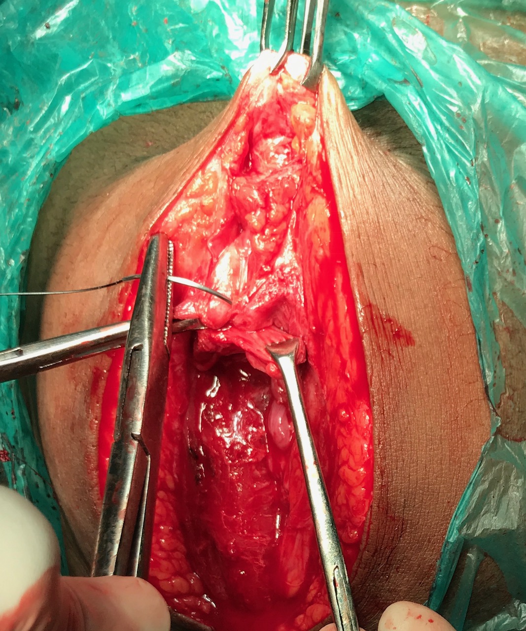
She was posted for the procedure after taking a written, informed consent. Intra-operative findings showed a 2 x 1 x 1 cm endometriotic mass overlying the rectus sheath and adherent to it. Wide excision of the tissue with surrounding clear margins was done and the specimen was sent for histopathological examination. The abdominal wall gap was reconstructed in layers. The histopathology report confirmed the diagnosis of scar endometriosis. She was followed up for a period of 3 months with no recurrence of symptoms.


Figure 1. Endometrial glands and stroma in scar tissue on microscopy (arrow)

Discussion

Endometriosis is a chronic, estrogen dependent gynecologic disorder. Although benign in nature, it has many features of malignancy like local invasiveness and the ability to spread to distant organs. A review of literature places the estimated incidence of cesarean scar endometriosis to be around 0.2 to 0.45 %.[3]
The pathogenesis of cesarean scar endometriosis involves iatrogenic implantation of endometrial cells into the site of surgery. These cells then proliferate and form a swelling, under the influence of female hormones and nourished by a rich blood supply. Risk factors known to be associated with the condition include a past history of obstetric surgeries, especially hysterotomy done in the late second to early third trimester of pregnancy.
The common complaints at presentation are swelling in and around the scar site, associated with continuous or cyclical pain, an increase in the swelling during menses and rarely, brownish discharge from the mass. Diagnosis is made based on the history and local examination. Ultrasonography with color Doppler is the only imaging tool needed and is the most cost effective.
Computed tomography or magnetic resonance imaging may be needed only to exclude other diagnoses and to define the extent of the lesion and its involvement with adjacent structures before the surgery. Desmoid tumor, scar granuloma, hematoma, neuroma and metastatic implants are some of the differential diagnoses for scar endometriosis.[5] When these need to be ruled out, FNAC may be a useful tool and it provides a preoperative tissue diagnosis.
The treatment of choice for scar endometriosis is surgery, the procedure being wide en bloc excision with clear margins.[6] Other management options include the use of drugs such as progestogens, combined oral contraceptive pills and synthetic steroids namely, danazol. These medications provide only temporary relief and the condition is known to recur after their discontinuation. Endometriosis can recur in the same site with incomplete excision, hence, wide excision should be ensured. Surgery also prevents conversion to malignancy, which is a rare complication.
For the prevention of scar endometriosis, certain surgical practices have been recommended. These include closure of the visceral and parietal peritoneum, washing the pelvic cavity with saline prior to closure and using separate instruments and suture materials for closure of the abdominal wall.[7] The usefulness of such practices is yet to be proved by studies and further research is needed to establish their effectiveness.

Conclusion

Cesarean scar endometriosis is a rare postoperative complication which should be considered as a differential diagnosis for any swelling arising in and around the scar site. Surgical practices to prevent iatrogenic transplantation of endometriotic tissue to the abdominal wall need to be explored further. 

References
  1. Viganò P, Parazzini F, Somigliana E, Vercellini P. Endometriosis: epidemiology and aetiological factors. Best Pract Res Clin Obstet Gynaecol. 2004; 18(2):177–200. 
  2. Zhang J, Liu X. Clinicopathological features of endometriosis in abdominal wall—clinical analysis of 151 cases. Clinical and Experimental Obstetrics and Gynecology. 2016; 43(3): 379–383.
  3. Khachani I, Adib AF, Bezad R. Cesarean Scar Endometriosis: An Uncommon Surgical Complication on the Rise? Case Report and Literature Review.  Case Reports in Obstetrics and Gynecology. 2017; 2017:8062924
  4. Uzunçakmak C, Güldaş A, Özçam H, Dinç K. Scar Endometriosis: A Case Report of This Uncommon Entity and Review of the Literature. Case Rep Obstet Gynecol. 2013; 2013: 386783. 
  5. Nominato NS, Prates LFVS, Lauar I, Morais J, Maia L, Geber S. Caesarean section greatly increases risk of scar endometriosis,” European Journal of Obstetrics Gynecology and Reproductive Biology. 2010;152(1):83–85.
  6. Sumathy S,  Mangalakanthi J, Purushothaman K, Sharma D, Remadevi C, Sreedhar S. Symptomatology and Surgical Perspective of Scar Endometriosis: A Case Series of 16 Women. J Obstet Gynaecol India. 2017;67(3):218-223. 
  7. Yela DA, Trigo L, Benetti-Pinto CL. Evaluation of Cases of Abdominal Wall Endometriosis at Universidade Estadual de Campinas in a period of 10 Years. Rev Bras Ginecol Obstet. 2017;39(8):403-407.
Citation

Kalappa SB, Tiwari N, Kolhe A, Chauhan AR.  Cesarean Scar Endometriosis- A Postoperative Complication On The Rise. JPGO 2017. Volume 4 No.11. Available from: http://www.jpgo.org/2017/11/cesarean-scar-endometriosis.html