Volume 4 Issue 9, September 2017

Editorial
Chauhan AR

True Umbilical Knot With a Live Baby
Oraekwe OI, Egbo BI, Kalu SA, Nduka EC.

Pregnancy In Left Horn Of Bicornuate Uterus
Thunga C, More V, Hatkar PA, Satia MN.

Severe hypothyroidism causing minimal change nephropathy in early pregnancy
Bora A, Madhva Prasad S, Gupta AS.

Adnexal Torsion Leading To Ovarian Cyst Rupture With Hemoperitoneum
Thakare R, Pardeshi S, Gupta AS.

Successful Pregnancy Outcome In A Case Of Cardiac Ischemia And Previous Uterine Perforation
Daruwale R, Tiwari N, Chauhan AR.

Cervical Atresia- A Difficult Diagnosis
Mehta N, Parulekar SV.

Inside Out Transobturator Tape Insertion Using Shirodkar’s Sling Needles
Parulekar SV

Uterine Rupture With Lung Metastasis In A Case Of Choriocarcinoma - A Rare Entity
Chavan V,  Sanghvi N, Shah NH, Paranjpe SH.

Preeclampsia In A Patient Of Hemiplegic Migraine
Parihar AS, Warke HS, Mayadeo NM.

Editorial

Chauhan AR

As obstetricians, we are only somewhat familiar with postpartum blues and depression, and when we encounter patients with severe symptoms, we usually refer them to a psychiatrist. In recent times, a more serious condition has been identified, i.e.  Postnatal or Postpartum Posttraumatic Stress Disorder (PP PTSD); this article aims to sensitize the reader to this disease entity.

Historically, from his experience with patients shocked by the events and wars of the French Revolution, Pinel wrote the first descriptions of war neuroses and described it as “cardiorespiratory neurosis” and acute stuporous posttraumatic state. Conflicts in World War I and II gave birth to terms such as “soldier's heart” and “shell shock”. However, it was the horrors of the Vietnam war and the mental anguish and “invisible wounds” that soldiers faced after returning home which made the medical profession more aware of PTSD and made this a household term. It was first mentioned in the 3rd edition of the Diagnostic and Statistical Manual of Mental Disorders (DSM-III) of the American Psychiatric Association in 1980, and as per the DSM -IV, it is classified under anxiety disorders. Though the incidence of PTSD is only 6 – 7 % among the average population following any traumatic stress, data from the U.S. Department of Veterans Affairs state that 13.8 % veterans of Iraq and Afghanistan wars suffer from PTSD and on an average, 22 veterans commit suicide every day.

Through the understanding of PTSD in war veterans, experts have identified that a similar process occurs following birth. PP PTSD is particularly seen in those patients who may have experienced a traumatic or difficult birth or even a perception of trauma during childbirth, history of rape or sexual assault in the past, or rape or intimate partner violence in the present pregnancy, or pregnancy complicated with cord prolapse, stillbirth, unplanned cesarean section or instrumental delivery, or baby requiring NICU care. Severe physical complication or injury like PPH, unexpected hysterectomy, perineal trauma, or high levels of medical intervention are also triggers. There is a complicated mix of objective and subjective factors, including feelings of loss of control, powerlessness, lack of privacy and dignity, and/or lack of communication, support and reassurance during the delivery, and poor postnatal care. Women with lack of social support, family history of psychiatric illness especially anxiety disorders, poverty, abuse, or childhood behavioural problems are more at risk.

The diagnosis of PTSD is important as misdiagnosis is common. Though there is some symptom overlap between postnatal depression and postnatal PTSD, the two conditions are different. Women are often misdiagnosed with depression, and therefore treatments are unsuccessful. However, women with PTSD may also have depression, but the presence of one does not always imply the presence of the other. It is possible that up to 25% of women who have PTSD remain undetected. Hallmarks of PP PTSD are nightmares, poor sleep, anger, numbness or sadness and avoidance of groups.

As per DSM -IV, diagnosis of PTSD is based on a cluster of symptoms, categorized from A to F. Criterion “A” is the stressor or the traumatic event, where the patient has experienced an event that involves actual or threatened death or serious injury, and her response involves intense fear, helplessness, or horror. “B” is re-experiencing or intrusions, where the traumatic event is re-experienced in the form of recurrent images, thoughts, perceptions, or distressing dreams of the event, acting or feeling as if the event was recurring, or intense psychological distress. “C” is persistent avoidance of stimuli and numbing, indicated by avoiding thoughts, feelings, or discussion, activities, places or people that bring back recollections; inability to recall; diminished interest, or feeling detached or estranged. Criterion “D” is increased arousal manifest by difficulty in falling asleep, irritability, difficulty in concentrating, exaggerated startle response. Criterion “D” also includes negative alteration in cognition and mood, such as memory problems exclusive to the event, distorted sense of blame, horror or sadness, and feelings of isolation or detachment from other people. Criterion “E” defines that the duration of symptoms should be for more than 1 month, while criteria “F”, “G” and “H” all describe the severity of the symptoms. To clinch the diagnosis of PTSD, at least one symptom each from criterion B and C, 2 each from increased arousal (D) and negative alteration in cognition (D) should be present, along with symptoms for at least a month (E). The differential diagnosis of PTSD is other anxiety disorders, OCD, panic disorder, substance-induced disorder, borderline personality disorder or malingering.

The treatment of PTSD requires multiple modalities, including initial education and support, and referrals for psychotherapy and pharmacotherapy. The starting point may be to review and discuss the labor notes; if symptoms are of less than 4 weeks duration, 2 out of 3 patients will not require further intervention except close observation.
Psychotherapy is the mainstay of treatment, and is aimed at crisis intervention, trauma-focused psychotherapy and systematic desensitization; hospitalization may sometimes be necessary. First line psychotherapies include Cognitive Behavioral Therapy (CBT) and Eye Movement Desensitization and Reprocessing (EMDR). CBT aims to build new cognitive skills and engage in new behaviors, or change existing ones. It includes Prolonged Exposure (PE) wherein the patient confronts situations that she has been avoiding until her distress decreases, and Cognitive Processing Therapy (CPT) wherein she examines and challenges thoughts about the trauma until she can change the way she feels. The EMDR process is based on REM sleep, where through eye movements, the brain processes disturbing images and eventually resolves them.

Evidence-based pharmacological treatments include the use of Selective Serotonin Reuptake Inhibitors (SSRIs); Paroxetine is the gold standard. Other agents like Serotonin-Norepinephrine Reuptake Inhibitors (SNRIs), benzodiazepines, antidepressants and mood stabilizers have also been used with limited efficacy. Duration of treatment is typically 8 to 12 weeks, with maintenance for at least a year. Other therapies like hypnotherapy, acupuncture, yoga, meditation, writing, running, have also been suggested, along with family support.

The long-term implications of PTSD involve greater risk of other psychiatric disorders, higher rate of unemployment, relationship problems and overall poorer quality of life. Hence as obstetricians, it is our responsibility to help prevent PTSD by offering a positive pregnancy experience. Our role is to provide education to patients about labor and delivery; offer information on best practices so as to prepare patients for pain, labor analgesia, episiotomy, instrumentation, CS; draw up and document a birth plan; document all events in labor room meticulously and be open and honest to audit/ questions postpartum. If symptoms of PTSD do occur, we should not ignore or treat them casually, but refer these cases early to appropriate facilities.

True Umbilical Knot With a Live Baby

Author Information

Oraekwe OI*, Egbo BI*, Kalu SA**, Nduka EC***.
(* Senior Registrar, ** Registrar, *** Head of Department. Department of Obstetrics and Gynaecology, Federal Medical Centre, Umuahia, Abia State, Nigeria.)

Abstract 

True umbilical cord knot is an uncommon form of umbilical cord accident. It carries a significant risk of adverse perinatal outcome. We present a case of a true umbilical knot in a 30 year old booked primigravida who had an emergency cesarean section on account of intrapartum fetal heart rate abnormality. Antenatal diagnosis of true knot remains a challenge especially in resource poor settings.

Introduction

True umbilical cord knot is a rare but important cause of perinatal morbidity and mortality. It affects 0.3-2.1 % of all pregnancies,[1-3] with a perinatal mortality rate as high as 11%.[1] True knots form when the fetus rotates in utero which occurs mostly between 9 and 28 weeks of gestation.[1] Antenatal diagnosis of a true knot remains a challenge because there is no specific clinical feature attributed to the condition.[4] Obstetric complications associated with this condition include cord compromise, fetal acidosis, low Apgar score at the first minute, higher risk of cesarean delivery and intrauterine fetal death.[5] Despite the consequences, the prenatal management of this condition is yet to be determined.[5]
                                                              
Case Report

A 30 year old primigravida, booked for antenatal care at Federal Medical Center, Umuahia at a gestational age (GA) of 12 weeks, on 20-12-2016. The index pregnancy was desired and spontaneously conceived. The booking weight was 64 kg; height was 1.64 m; blood pressure was 120/80 mm of Hg; hemoglobin concentration was 11.3 g/ dl, and urinalysis showed no abnormality.
Her hemoglobin genotype is AA and blood group is O rhesus D positive. The venereal disease research laboratory test was nonreactive and she tested negative to human immunodeficiency virus I and II. The index pregnancy remained unremarkable and ultrasonography done at 24 weeks GA revealed normal findings. Color Doppler ultrasonography was not done because it was not a routine antenatal investigation in the hospital. She was not a known hypertensive or a diabetic. Following the onset of labor pain on 26-06-2017 at a GA of 39 weeks, examination revealed a young lady in painful distress, afebrile, not pale and anicteric. There was no pedal edema. The pulse rate was 90 beats per minute (bpm), full volume and regular. The blood pressure was 120/80 mm of Hg. The heart sounds I and II were heard and there was no murmur. The respiratory rate was 24 cycles per minute and the breath sound was vesicular. The abdomen was enlarged and moved with respiration. The symphysio-fundal height was 38 cm. The lie was longitudinal and presentation was cephalic with the head 5/5 palpable per abdomen. The fetal heart rate was 164 bpm. Vaginal examination revealed cervical dilatation of 5 cm with the vertex at station 0-3. There was no cord prolapse. She was admitted into the labor ward where immediate intrauterine fetal resuscitation was commenced using intranasal oxygen at 6 liters per minute, intravenous normal saline at 30 drops per minute and she was placed on left lateral position. About an hour later the fetal heart rate however remained persistently elevated ranging from 168 to 175 bpm, necessitating an emergency cesarean section. Outcome was a male neonate which weighed 3.2 kg, with an Apgar score 9 and 10 in the first and fifth minute respectively. There was no retroplacental clot. The umbilical cord length was 70 cm with no gangrene. There was no gross fetal abnormality. The estimated blood loss was 400 ml. A true knot was found 40 cm from the placental insertion of the cord (Figure 1). The postoperative period was unremarkable and the woman was subsequently discharged on the 5th postoperative day.


Figure 1. The umbilical cord with a true knot.

Discussion

The umbilical cord, which is an essential connection between the mother and the fetus, provides the baby with essential metabolites while transferring waste products back to the mother. Occasionally the cord is prone to a number of intrinsic or accidental disorders which may disrupt the cord morphology, decrease the blood flow in the cord and ultimately result in fetal compromise.[6] There are about 30 conditions which can result in cord accidents and true umbilical knot is one such.[1] Risk factors for this condition include excessively long cord, polyhydramnios, monoamniotic twin gestation, male fetuses, amniocentesis, obesity, gestational diabetes mellitus, advanced maternal age, small for gestational age fetus and multiparity.[5,7] The male baby was the only predisposing factor noted in our case. 
In the antenatal period, the suspicion of a true knot can be entertained with difficulty by visualizing the ‘hanging noose sign’ on a gray-scale ultrasonography, however, the confirmation requires a color Doppler three-dimensional view.[1,4] This difficulty was shown by normal ultrasonographic findings noted in our case.
Most true knots are loose with no adverse pregnancy outcome except when they are tight,[8] and this rarely occurs prior to the onset of labor.[4] Our case had uneventful antenatal period, however, with onset of labor the true knot probably became tight manifesting as persistent fetal heart tachycardia. The good fetal outcome was due to immediate intrauterine resuscitation and timely abdominal delivery.
True umbilical knot is a rare but important cause of perinatal death. Even though its antenatal diagnosis has been advocated, this raises the question whether the women with the diagnosis should be allowed to labor or not. However for women with diagnosis of fetal heart rate abnormality in labor, true umbilical knot should be entertained as a possible cause and timely intervention may avert fetal demise.

References
  1. Kumar SS, Priya BA. True umbilical cord knot. Indian Journal of Health Sciences and Biomedical Research KLEU 2015; 8(2): 136-8.  
  2. Kotingo EL, Allagoa DO. True umbilical cord knot, nochal cord and cord round body with favourable obstetric outcome in an unbooked elderly nullipara: a case report and literature review. International Journal for Research in Health Sciences and Nursing 2016; 2(2): 25-31.
  3. Airas U, Heinonen S. Clinical significance of true umbilical knots: a population-based analysis. Am J Perinatol. 2002;19(3):127-32. 
  4. Bohîlțea RE, Turcan N, Cîrstoiu M. Prenatal ultrasound diagnosis and pregnancy outcome of umbilical cord knot-debate regarding ethical aspects of a series of cases. Journal of Medicine and Life 2016; 9(3): 297-301. 
  5. Räisänen S, Georgiadis L, Harju M, Keski-Nisula L, Heinonen S. True umbilical cord knot and obstetric outcome. International Journal of Gynecology and Obstetrics 2013; 122(1): 18-21. 
  6. Bangal VB, Shinde KK, Gavhane SP, Borawake SK , Chandaliya RM. Twisting of the umbilical cord causing intrauterine fetal death. International Journal of Biomedical and Advance Research IJBAR 2012; 3(8): 656-9.
  7. Khan M, Zahiruddin S, Iftikhar M.  True knot of umbilical cord: case report and review of literat. J Pak Med Assoc 2016; 66(8): 1037-8.
  8. Szczepanik ME, Wittich AC. True knot of the umbilical cord: a report of 13 cases. Military Medicine 2007; 172(8): 892-4.
Citation

Oraekwe OI, Egbo BI, Kalu SA, Nduka EC. True Umbilical Knot With a Live Baby. JPGO 2017. Volume 4 No.9. Available from: http://www.jpgo.org/2017/09/true-umbilical-knot-with-live-baby.html

Pregnancy In Left Horn Of Bicornuate Uterus

Author Information

Thunga C*, More V**, Hatkar PA***, Satia MN****
(* Senior Resident, ** Assistant Professor, *** Associate Professor, **** Professor, Department of Obstetrics and Gynecology, Seth GS Medical College and KEM Hospital, Mumbai, India.)

Abstract 

We report a case of bicornuate uterus with pregnancy in the left horn with an unusual presentation. It was diagnosed at 14 weeks of pregnancy. Patient was referred from a peripheral private nursing home with failed attempts at surgical evacuation for missed abortion. At our hospital, exploratory laparotomy with excision of the left gravid horn of the uterus was done.

Introduction

Congenital malformations of female genital tract result from embryologic maldevelopment of the mullerian ducts either in their formation, fusion or absorption stage. They are the deviations from normal anatomy, representing a benign condition with a prevalence of 5.5% in general population.[1] Bicornuate uterus results from incomplete fusion of the two mullerian ducts leading to varying degrees of separation between two symmetrical uterine cavities ranging from partial separation to complete septation with no communication between the two cavities.[2] Bicornuate uterus is known to be associated with several obstetric complications; some of them include recurrent pregnancy loss, fetal malpresentation, intrauterine growth restriction, preterm labor, increased need for operational intervention including cesarean section.[3] Among these, the most common ones affecting the outcome of pregnancy are recurrent pregnancy loss (25%), preterm birth (15-25%) and cervical insufficiency (38%).[4] We are reporting one such case of bicornuate (bicornis unicollis) uterus with pregnancy in one horn. 

Case Report

A 20 year old, married since 2 years, gravida 2, abortion 1 with 13.3 weeks missed abortion on ultrasonography (USG), was referred from a private hospital in view of bicornuate uterus with failed repeated attempts at evacuation. She presented to a private consultant obstetrician with spotting per vaginum. Ultrasonography had showed a missed abortion of 13.3 weeks in the left horn of a bicornuate uterus with marginal placenta previa. Right side horn showed thickened endometrium. She was treated with tablet Misoprostol 400 microgram per vagina, followed by injection oxytocin 10 units intravenously. In view of failed attempt, she went to another private consultant where ultrasound guided aspirotomy was attempted but dilator invariably went into the non pregnant horn. Hence the procedure was abandoned and she was referred to our tertiary care center for further management.
She came to emergency receiving room 4 days later with complaints of abdominal pain and vaginal bleeding since past 4 days, and soakage of almost 4 pads per day. She also had complaints of giddiness since that morning. She had no significant past medical or surgical illness. Her menstrual history was normal with regular cycles previously. Her obstetric history revealed a previous spontaneous abortion at 3 months, 1 year back after which curettage was not done. Present pregnancy was a spontaneous conception.  
Her vital parameters were within normal limit. General condition was fair. She was conscious, well oriented in time and space, and afebrile. Mild pallor was noted. Her pulse was 96 beats per minute with good volume, blood pressure was 110/70 mm of Hg. Respiratory and cardiovascular system examination were normal. On abdominal examination uterus was 14 weeks in size, and lower abdominal tenderness was noted. There was no guarding or rigidity. On speculum examination cervix, and vagina were healthy. Minimum bleeding through os was present. On digital vaginal examination external os admitted tip of finger, and internal os was closed. Uterus was 14 weeks in size.
Biochemical and serological tests showed a Hb of 8.9 gm%, WBC count of 11,800 /cumm, platelet count of 3.08 lakhs/cumm. Renal and liver function tests were within normal limits. Coagulation profile was within normal limits. Injectable antibiotics were started. Ultrasound pelvis showed missed abortion of 14 weeks in the left horn of a bicornuate uterus. Contrast enhanced CT pelvis reported a bicornuate uterus with gestational sac in left uterine cornua measuring 8.8x7.5 cms with fetal parts seen within. Exploratory laparotomy was planned electively. Intraoperatively, 15x15 cms sized left side horn of bicornuate uterus with pregnancy in situ was noted, uterine wall was thinned out and stretched with bluish products of conception. The right side horn of uterus that measured 6x5x4 cms was externally continuous with the left horn. The pregnant left horn communicated with only the right horn and had no communication with the single cervical canal as shown in the image (Figure1). Bilateral fallopian tubes and ovaries were normal. Excision of the left pregnant horn with repair of the uterine defect was done. Intra operatively she was given one unit of packed red cell transfusion as her pre-operative hemoglobin was 8 g%. She remained stable after surgery. Wound check was done and she was discharged with advice to use contraception for the next 6 months. Histopathology report confirmed the diagnosis of uterine horn with fetus and placenta.


Figure 1. Intra operative picture showing left horn with products of conception (yellow arrow), normal sized right horn of uterus (blue arrow), communication between the right and left horns (white thin arrow).


Figure 2. Excised left uterine horn with abortus (fetus with and the placenta) 

Discussion

Bicornuate uterus (bicornis unicollis) represents a double uterus with a single cervix and vagina resulting from the failure of the embryologic lateral fusion of the Mullerian ducts. Each uterus has a horn linked to its own fallopian tube and ovary. Bicornuate uterus is classified as class four according to the classification given by sub-committee of the American Fertility Society, now the American society of Reproductive Medicine.[5] According to the EAC (Embryological aberration, Anatomical abnormalities and Clinical features) classification of congenital malformations of the female genital tract, our case can be classified as UtR 6 cm Fn No Obs, UtL 15 cm Fn No Obs. That is, bicornuate uterus with right horn of 6 cms, functional and no obstruction, left horn of 15 cms, functional and no obstruction.[6] The pregnant left horn communicated with only the right horn and had no communication with the single cervical canal as shown in Figure1.  Uterine anomalies during pregnancy may be difficult to diagnose only by two-dimensional ultrasonography.[7] Recently three dimensional ultrasonography has been introduced as a non invasive diagnostic modality with good spatial awareness, hence it is considered to be superior to other modalities.[8] The incidence of rupture of the malformed uterus is 8-11 %.[9,10] This is due to inability of the malformed uterus to expand as pregnancy advances. In our case, the chance of pregnancy growing had it been live would be very little as already there was thinning of uterine wall. She was probably fortunate that the horn did not rupture as pregnancy ceased to grow. However, there have been several reports of varied clinical presentations of pregnancy in a bicornuate uterus in literature including rupture of the pregnant horn as well as successful outcomes with live pregnancy reaching upto term.[11,12] An ultrasonography early in pregnancy helps in diagnosing the uterine anomalies. Traditional treatment includes exploratory laparotomy with excision of the pregnant horn to prevent rupture and recurrence of the condition. A high index of suspicion is necessary to diagnose a case of Mullerian anomaly whenever we encounter cases that do not respond to induction of labor. Thereby catastrophic events like rupture of the pregnant horn and maternal mortality can be avoided. 

References
  1. Chan YY,  Jayaprakasan K, Tan A, Thornton JG, Coomarasamy A, Raine-Fenning NJ. Reproductive outcomes in women with congenital uterine anomalies: a systematic review. Ultrasound Obstet Gynecol 2011; 38(4): 371–382.
  2. Reichman DE, Laufer MR. Congenital uterine anomalies affecting reproduction. Best Pract Res Clin Obstet Gynecol 2010; 24(2): 193–208.
  3. Gogineni S, Devi KJP, Vellanki VS, Sravanthi M, Dharmaja D. Bicornuate uterus with successful pregnancy outcome. IOSR journal of dental and medical sciences 2013; 9(3):52-54.
  4. Lin PC. Reproductive Outcomes in Women with Uterine Anomalies. J Women Health (Larchmt) 2004; 13(1): 33-39.
  5. The American Fertility Society classifications of adnexal adhesions, distal tubal occlusion, tubal occlusion secondary to tubal ligation, tubal pregnancies, müllerian anomalies and intrauterine adhesions. Fertil Steril. 1988;49(6):944-55.
  6. Parulekar SV. EAC Classification Of Congenital Malformations Of The Female Genital Tract. JPGO 2015; 2(4) 
  7. Hua M, Odibo AO, Longman RE, Macones GA, Roehl KA, Cahill AG. Congenital uterine anomalies and adverse pregnancy outcomes. Am J Obstet Gynecol 2011; 205(6): 558.e1-5
  8. Ahmadi F, Zafarani F, Haghighi H, Niknejadi M, Dizaj AVT. Application of 3D Ultrasonography in Detection of Uterine Abnormalities. Int J Fertil Steril 2011;4(4): 144-147
  9. Walsh CA, Baxi LV. Rupture of the primigravid uterus: a review of the literature. Obstet Gynecol Surv. 2007;62(5):327-34
  10. Ravasia DJ, Brain PH, Pollard JK. Incidence of uterine rupture among women with müllerian duct anomalies who attempt vaginal birth after cesarean delivery. Am J Obstet Gynecol 1999;181(4):877–81
  11. Kore S, Pandole A, Akolekar R, Vaidya N, Ambiye VR. Rupture of left horn of bicornuate uterus at twenty weeks of gestation. J Postgrad Med. 2000;46(1):39-40.
  12. Adeyemi AS, Atanda OO, Adekunle AD. Successful pregnancy in one horn of a bicornuate uterus. Ann Afr Med. 2013;12(4):252-4.
Citation

Thunga C, More V, Hatkar PA, Satia MN. Pregnancy In Left Horn Of Bicornuate Uterus.  JPGO 2017. Volume 4 No.9. Available from: http://www.jpgo.org/2017/08/pregnancy-in-left-horn-of-bicornuate.html

Severe Hypothyroidism Causing Minimal Change Nephropathy In Early Pregnancy

Author Information 

Bora A*, Madhva Prasad S**, Gupta AS***

(* Second Year Resident, ** Assistant Professor, *** Professor, Department of Obstetrics and Gynecology, Seth GS Medical College and KEM Hospital, Mumbai.)

Abstract

Vulval edema, as a part of pregnancy is a common entity. While hypertension and renal disease are the commonly attributed etiologies, these may be transient also. Here a case of severe hypothyroidism at 16 weeks pregnancy which presented as a transient nephrotic syndrome is described. Complete resolution with correction of thyroid status occurred.  

Introduction 

Occurrence of vulval edema, generalized edema all over the body, high blood pressure and proteinuria in pregnancy usually signifies preeclamptic state. However in this case, the occurrence of proteinuria was probably because of a minimal change nephropathy which can occur in severe hypothyroidism.

Case Report 

A 24 year old primigravida with 16 weeks pregnancy presented with swelling all over the body and decreased urine output. She was antenatally unregistered. She noticed gradual swelling, initially in the feet and vulvar region, around three weeks prior, which gradually progressed to involve entire body. She gave no history of burning micturition, dysuria or increased frequency of micturition or urgency. She had no complaints of breathlessness, palpitations or weakness. She gave no complaints of yellowish discoloration of the body, pruritus, clay colored stools or pain in abdomen.
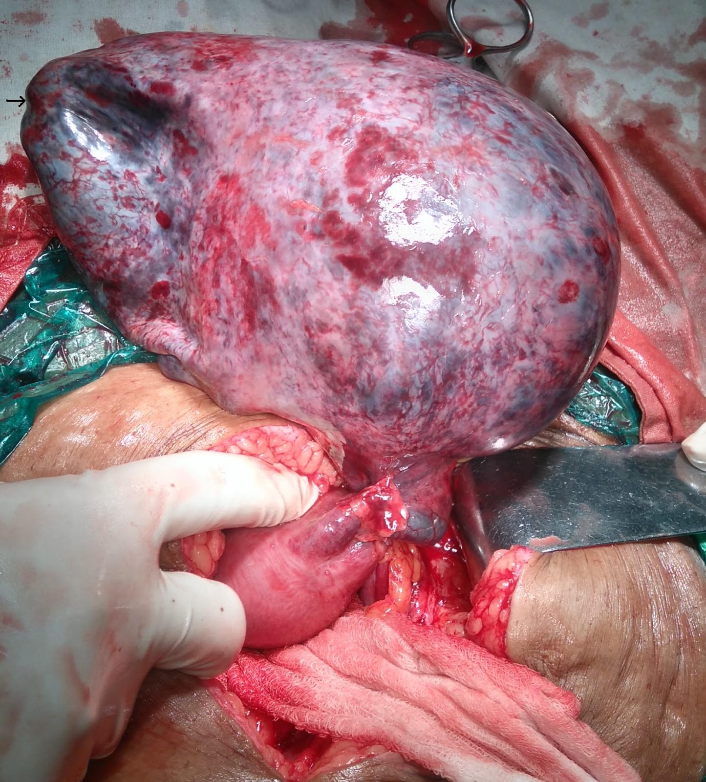
On examination her blood pressure and pulse were 130/80 mm of Hg and 88 beats per minute respectively. Cardiovascular examination was within normal limits and chest was clear. Bilateral non-pitting pedal edema was present. Abdomen was soft, free fluid was detected and uterus was just palpable. There was gross vulvar edema. On per vaginal examination internal os was closed and uterus was 14 weeks in size. 
Ultrasonography showed a live intrauterine gestation of 14 weeks 6 days, bilateral mild pleural effusion and ascites. She was admitted for evaluation of anasarca. Hemoglobin was 8.6 gm %, white blood cell count was 7600 cells/ cubic mm, platelet count was 3.6 lacs, serum creatinine was 0.7 mg%, blood urea nitrogen was 5.1 mg%, sodium and potassium levels were 143 meq/ l and 4.2 meq/ l respectively. Liver function tests, including serum protein levels were within normal limits.   
Urine routine microscopy was within normal limits, except a trace of albumin. Serum TSH was done, and surprisingly was found to be extremely elevated; 150 mg/dl. The other abnormal investigation was twenty four hour urine protein which was found to be high; 78.7mg/ dl (normal < 10mg/dl). Nephrologist advised to rule out autoimmune causes. Anti-nuclear antibodies and anti dsDNA antibodies, ICT/ DCT were negative; C3 and C4 were within normal limits.  
She was started on oral levothyroxine 150 mcg once a day. During her stay in the hospital, strict blood pressure monitoring was done. In view of a single high blood pressure value (150/100 mm Hg) and in the setting of proteinuria she was started on methyldopa 250 mg BD, and labetalol 50 mg BD; since the nephrologists anticipated rise in blood pressure with progression of pregnancy. For the vulvar edema, regular magnesium sulphate dressings were done. Fundoscopy was done and retina was found to be normal. Ultrasonography for kidney, ureter and bladder was normal. She was discharged with the advise to continue all the above 3 drugs and follow up as an outpatient. However, she presented to the outpatient department only after 2 months.
In her consequent visits, she had no facial puffiness, no pedal edema and the vulvar edema had significantly reduced. Nephrologist stopped the antihypertensive medications. She continued home blood pressure monitoring. Urine for albumin was always nil and obstetric Doppler was normal. She was admitted at 38 weeks for induction of labor for chronic hypertension. Critical review of her case history, her initial clinical presentation of anasarca, hypertension and its subsequent disappearance after starting thyroid replacement therapy enabled us to co-relate her clinical condition to severe hypothyroidism and exclude the differential diagnosis of minimal change nephrotic syndrome or chronic hypertension.  Induction of labor was deferred and she was allowed to go into spontaneous labor at 39 weeks and 4 days gestation. She remained normotensive during labor. She progressed spontaneously and delivered a female child of 2.438 kg, with an Apgar score of 9/10. Course of labor was uneventful. Repeat serum TSH value was 9.2 mg/ dl. She was continued on levothyroxine 150 microgram and discharged.

Discussion 

The occurrence of hypothyroidism ranges from around 3 to 15 % in various population groups.[1] Guidelines for management of hypothyroidism and screening for subclinical disease in pregnancy has now become universally accepted.[2] However, the clinical presentation of hypothyroidism can be varied. Our patient initially presented with gross vulval edema and ascites. Vulvar edema can be due to hypoproteinemia, and may be associated with hypertension in pregnancy.[3]
Hypothyroidism presenting as ascites is a rare but well established entity.[4] Though our patient had normal protein values, she had few readings of elevated blood pressure and anti-hypertensives were started though blood pressure did not reach the usually recommended values of 160/110mm Hg.[5]
However, in 2 months, the antihypertensives required discontinuation and patient did not have any further high blood pressure readings. Such a normotensive state continued throughout pregnancy, labor and her post-partum period. Hence, it was retrospectively considered that the initial presentation of edema was probably not due to the hypertension or related renal disease. De Feudis et al have described ascites as the initial presentation of primary hypothyroidism, albeit in a non-pregnant state.[6] Mizgala et al and Patel et al had also previously reported preeclamptic patients presenting with hypothyroidism.[7,8] However, now recent evidence has convincingly proved that hypothyroid disease is associated with hypertension in pregnancy.[9] This association may be genetically determined also, as reported recently by Procopciuc et al.[10]
Our patient had a TSH value of > 150 mg/dl, which is considered as severe hypothyroidism. This level was detected at the time of evaluation of anasarca during her initial presentation. Inversetti et al have also described a case of severe hypothyroidism which mimicked a preeclamptic state and highlighted the need for correction of thyroid status before any further intervention.[11] Ipadeola et al have also reported a case where hypothyroidism was unmasked by preeclampsia and ascites, almost identical to our case.[12]
The nephrologists were of the opinion that this presentation was similar to Minimal Change renal disease, though biopsy was not done. This is consistent with the case reported by Soh et al wherein minimal change nephrotic syndrome occurred with deterioration of hypothyroidism.[13] Karethimmaiah et al have also reported the need for increased thyroxine supplementation in patients with nephrotic syndrome.[14]
Hence it is retrospectively hypothesized that our patient developed a transient nephrotic syndrome like picture due to severe hypothyroidism, presenting as anasarca, that resolved completely when she achieved the euthyroid state. That a minimal change renal disease or chronic hypertension did not exist is proven by the fact that the patient went till term gestation with no further high blood pressure readings, proteinuria or intrauterine fetal growth retard or fetal compromise. However, this enlightenment occurred only at 38 weeks of gestation during a critical review of her clinical course when we were contemplating induction of labor for chronic hypertension. 
To conclude, hypothyroidism can present in a varied manner. This case report also highlights the fact that clinical decision making is an arduous task requiring correlation of many factors and in spite of superspecialists’ opinions, the judgment of a senior obstetrician is indispensable.  

References
  1. Negro R, Stagnaro-Green A. Diagnosis and management of subclinical hypothyroidism in pregnancy. BMJ. 2014;349:g4929.
  2. Alexander EK, Pearce EN, Brent GA, Brown RS, Chen H, Dosiou C, et al. 2017 Guidelines of the American Thyroid Association for the Diagnosis and Management of Thyroid Disease During Pregnancy and the Postpartum. Thyroid. 2017;27(3):315–89
  3. Daponte A, Skentou H, Dimopoulos KD, Kallitsaris A, Messinis IE. Massive vulvar edema in a woman with preeclampsia: a case report. J Reprod Med. 2007; 52(11):1067–9. 
  4. Philips CA, Sinha U, Chattopadhyay P, Mukhopadhyay P, Haldar S. Isolated ascites in hypothyroidism: medical and ethical issues. J Indian Med Assoc . 2010; 108(8):523–4. 
  5. Cunningham FG. Hypertenisve disorders. In Cunningham FG, Leveno KJ, Bloom SL, Spong CY, Dashe JS, Hoffman BS, editors. Williams’ Obstetrics. 24th ed. New York. Mc Graw hill 2014; pg 745-9
  6. De Feudis L, Scudieri M, Orlando D, Traisci G. Ascites as preeminent manifestation of primary hypothyroidism. Clinical case. Ann Ital Med Int.1999;14(4):294–7. 
  7. Mizgala L, Lao TT, Hannah ME. Hypothyroidism presenting as hypothermia following pre-eclampsia at 23 weeks gestation. Case report and review of the literature. Br J Obstet Gynaecol. 1991; 98(2):221–4
  8. Patel S, Robinson S, Bidgood RJ, Edmonds CJ. A pre-eclamptic-like syndrome associated with hypothyroidism during pregnancy. Q J Med. 1991; 79(289):435–41. 
  9. Wilson KL, Casey BM, McIntire DD, Halvorson LM, Cunningham FG. Subclinical Thyroid Disease and the Incidence of Hypertension in Pregnancy. Obstet Gynecol. 2012; 119 (2, Part 1):315–20. 
  10. Procopciuc LM, Caracostea G, Hazi G, Nemeti G, Stamatian F. D2-Thr92Ala, thyroid hormone levels and biochemical hypothyroidism in preeclampsia. Gynecol Endocrinol . 2017; 33(2):136–40. 
  11. Inversetti A, Serafini A, Manzoni MF, Dolcetta CA, Valsecchi L, Candiani M. Severe hypothyroidism causing pre-eclampsia-like syndrome. Case Rep Endocrinol. 2012;2012:586056. Available from: http://www.ncbi.nlm.nih.gov/pubmed/22937297 
  12. Ipadeola A, Nkwocha GC, Adeleye JO. Subclinical hypothyroidism unmasked by preeclampsia and ascites. Indian J Endocrinol Metab. 2013; 17(Suppl1): S173–S175.
  13. Soh S, Aki O, Manabu O, Norimasa K, Hiroshi K, Masao N. A case of minimal change nephrotic syndrome with hypothyroidism deterioration. CEN case reports.  2016;5(1):95–8. 
  14. Karethimmaiah H, Sarathi V. Nephrotic Syndrome Increases the Need for Levothyroxine Replacement in Patients with Hypothyroidism. J Clin Diagn Res; 2016;10(12):OC10-OC12
Citation

Bora A, Madhva Prasad S, Gupta AS. Severe hypothyroidism causing minimal change nephropathy in early pregnancy. JPGO 2017. Volume 4 No.9. Available from: http://www.jpgo.org/2017/08/severe-hypothyroidism-causing-minimal.html

Adnexal Torsion Leading To Ovarian Cyst Rupture With Hemoperitoneum

Author Information 

Thakare R*, Pardeshi S**, Gupta AS***

(* Second Year Resident, ** Assistant Professor, *** Professor, Department of Obstetrics and Gynecology, Seth GS Medical College and KEM Hospital, Mumbai, India.)

Abstract

A 35 year old woman came to our tertiary care hospital with pain in abdomen and vomiting since 3 days with  ultrasound (USG) scan suggestive of right ovarian mass of 16.6x11.6x 8.6 cm. CA 125 was 43 U/ml. Repeat USG done at our center was suggestive of large multiloculated lesion of 16x8x16 cm in right adnexa with thick septations within and large cystic components showing internal echoes. CT scan (pelvis) was suggestive of ovarian neoplasm with torsion. Emergency exploratory laparotomy was done. Intra-operatively a 17x17 cm ovarian cyst on right side with torsion at level of right cornua was encountered. Pedicle had two twists. Right fallopian tube and ovarian cyst were gangrenous with hemorrhagic fluid inside. Necrosed apex of the cyst had ruptured causing a hemoperitoneum.  Right sided salpingo-oophorectomy was done. 

Introduction

Ovarian torsion is one of the most common gynecological emergencies and a significant cause of acute lower abdominal pain in women of all age groups. The ovary along with its ipsilateral fallopian tube is typically involved. Ovarian torsion commonly occurs in a pathologically enlarged ovary. It is mostly unilateral. The torsion can involve the ovary alone or both the ovary and the fallopian tube (adnexal torsion). The right sided adnexa is more commonly involved (60%).

Case Report

A 35 year old woman married since 8 years was referred from a private hospital with pain in abdomen and vomiting since 3 days with outside ultrasound (USG) scan suggestive of right ovarian mass of 16.6x11.6x8.6 cm. CA 125 was 43U/ml.
On admission, she was afebrile, pulse was regular, and good volume. Her blood pressure was normal. Systemic examination was normal. On abdominal examination, abdomen was soft with minimal guarding, a mass of about 26 weeks size was felt more on the right side of the midline and it was tender. Rigidity was absent. On per speculum examination minimal discharge was present. On per vaginal examination vagina was warm with tenderness in the right fornix. Cervical motion tenderness was present. Mass was felt to be arising from the right side and uterus was felt separately behind the mass.
Repeat USG done was suggestive of a large multiloculated solid cystic lesion 16x8x16 cm in the right adnexa with thick septations within and large cystic components showing moving internal echoes; largest cyst measuring 9x8 cm with no evidence of vascularity. Left ovary was normal. CT scan (pelvis) was suggestive of a well defined solid cystic lesion 16x12x13 cm with areas of necrosis in the right adnexa extending upto the right lumbar region with loss of fat planes. Features were suggestive of ovarian neoplasm with torsion. (Figure 1)
She had one episode of fever of 38.6º F. Serological and biochemical tests were negative except for elevated leucocyte count of 19,500 cells/ml with 80 % neutrophils. Prophylactic antibiotics were started.


Figure 1. CT scan pelvis.

Emergency exploratory laparotomy was done. Intra-operatively a 17x17 cm ovarian cyst on the right side with torsion at level of right cornua was encounterd. It had two twists. (Figure 2). Right fallopian tube and ovarian cyst were gangrenous with hemorrhagic fluid inside (Figure 3,4). Necrosed apex of cyst had ruptured causing hemoperitoneum of around 300 ml. Right sided salpingo-oophorectomy done.  Right sided cystic fluid was sent for cytology and specimen was sent for histopathological examination. Postoperative period was uneventful. Histopathological examination studied from the ovarian tissue and fallopian tube showed extensive hemorrhagic necrosis, no viable tissue, features suggestive of torsion.


Figure 2. Right Ovarian cyst  with torsion at level of right cornua (arrow).


Figure 3.  Necrosed apex of the cyst (arrow)


Figure 4. Necrosed apex of cyst leading to hemoperitoneum (area between the two Babcock’s forceps)

Discussion

The occurrence of ovarian cysts varies from 5 to 15%. Complications such as rupture, torsion (15%) and rarely malignant changes (2%) may be seen. In adnexal torsion ovary and the fallopian tube undergo twisting. The ovary and the fallopian tube may rotate as a single entity in most of these cases.[1] About 3% of gynecologic emergencies are attributed to adnexal torsion and occur most commonly in women of reproductive age group. The other types of  torsion commonly seen are only ovarian and torsion of isolated fallopian tube. The right sided adnexa is more commonly involved,  mostly because of the restricted mobility of left ovary by the sigmoid colon.  The conditions with increased mobility of adnexal masses which predispose them to torsion are congenitally long utero-ovarian ligament and large ovaries (diameter > 6 cm).[2] The adnexal torsions are divided into 3 categories that are acute (< 24 hours), subacute (2 days) and chronic (> 3 days) depending on the time duration between onset of symptoms and surgical management.  
In torsion compression of veins occurs followed by that of arteries leading to infarction and necrosis. The sequence of events leads to friable tissue which is prone for rupture. Hemorrhage in the cyst with subsequent rupture resulting into hemoperitoneum may occur especailly when diagnosis or treatment or both are delayed.
Subacute torsion with cyst rupture and hemoperitoneum presents with dull aching pain in lower abdomen and leucocytosis (as seen in our case) and subsequently hemorrhagic shock.[3]  The common causes of ovarian cyst rupture are idiopathic, torsion, malignancy, trauma, anticoagulation therapy and iatrogenic cause. The ovarian cyst rupture with minimal hemorrhage is mostly physiological. Pathological cysts are serous and mucinous cystadenomas, dermoid cysts and endometriotic cyst.
The initial investigation of choice is ultrasound (USG) in which twisted pedicle has been described as a bull’s eye target, whirlpool or snail shell.[4] Transvaginal color doppler sonography often shows disruption of normal adnexal blood flow. USG  is also the preferred initial investigation for suspected ovarian cyst hemorrhage or rupture. The findings are of an enlarged ovary containing bizarre mixed echoes in a reticular pattern like a spider’s web.  The blood clot within the cyst may ‘wobble’ in a jelly-like fashion on pressing with the  transvaginal probe. If significant hemorrhage is suspected or the cause is unclear, contrast-enhanced CT scan is often the investigation of choice.
Management includes either salvaging the adnexa or resection of associated cyst or tumor and oophoropexy. In cases presenting with hemoperitoneum stabilization may be required with blood and blood products transfusion. Removal of adnexal structures may be required in cases with necrosis or rupture. Detorsion of the involved adnexa can be attempted in early cases of torsion to salvage the ovary. The lesions of the ovary like ovarian cyst may be dealt with by cystectomy and ovarian reconstruction. Unilateral salpingo-ophorectomy is the most common surgical procedure done for adnexal torsion.[5]  Laparoscopy or  laparotomy can be done for surgical management of an adnexal mass. The surgical approach is chosen based on the degree of suspicion of malignancy, patients preference and surgeons expertise. To prevent retorsion, unilateral or bilateral oophoropexy can be done. This includes shortening of the utero-ovarian ligament using a running stitch through the ligament or either the ovary or the utero-ovarian ligament is sutured  to the posterior aspect of the uterus, the round ligament or the lateral pelvic wall.[6]

Conclusion

If this patient had reported early, the gangrenous changes could have been prevented by early intervention there by salvaging the ovarian tissue. This case highlights the importance of early detection and intervention in suspected cases of ovarian torsion especially in women of reproductive age group.

References 
  1. McWilliams GD, Hill MJ, Dietrich CS 3rd. Gynecologic emergencies. Surg Clin North Am 2008;88(2):265-83.
  2. Hoffman BL. Pelvic mass. In Hoffman BL, Schorge JO, Schaffer JI, Halvorson LM, Bradshaw KD, Cunningham FG.editors Williams Gynecology 2nd edition. Texas: Mc Graw Hill Medical 2012;  pp. 246-280.
  3. Bottomley C, Bourne T. Diagnosis and management of ovarian cyst accidents. Best Pract Res Clin Obstet Gynaecol 2009;23(5):711–24.
  4. Vijayaraghavan SB. Sonographic whirlpool sign in ovarian torsion.J Ultrasound Med. 2004 Dec;23(12):1643-9
  5. Takeda A, Hayashi S, Teranishi Y,  Imoto S, Nakamura H. Chronic adnexal torsion: an under-recognised disease entity. Eur J Obstet Gynecol Reprod Biol. 2017 Mar;210:45-53. 
  6. Djavadian D, Braendle W, Jaenicke F: Laparoscopic oophoropexy for the treatment of recurrent torsion of the adnexa in pregnancy: case report and review. Fertil Steril 2004 82(4):933-6.
Citation

Thakare R, Pardeshi S, Gupta AS. Adnexal Torsion Leading To Ovarian Cyst Rupture With Hemoperitoneum.  JPGO 2017. Volume 4 No.9. Available from: http://www.jpgo.org/2017/09/adnexal-torsion-leading-to-ovarian-cyst.html

Successful Pregnancy Outcome In A Case Of Cardiac Ischemia And Previous Uterine Perforation

Author Information

Daruwale R*, Tiwari N**, Chauhan AR***.
(* Senior Registrar, ** Assistant Professor, *** Professor. Department of Obstetrics and Gynecology, Seth G. S. Medical College and K. E. M. Hospital, Mumbai, India.)

Abstract

Uterine perforation is an uncommon but potentially serious complication of uterine manipulation, evacuation of retained products of conception, termination of pregnancy, electroscopic procedures and during intra uterine device insertion. Termination of pregnancy is the most common procedure associated with uterine perforation. Pregnancies associated with previous uterine perforation have a higher morbidity rate in subsequent pregnancies. Myocardial ischemia is very rare in pregnancy but has been associated with poor maternal and fetal outcomes. We present a case of pregnancy complicated with cardiac ischemia and previous uterine perforation repair following suction evacuation, with successful maternal and fetal outcome.

Introduction

The incidence of uterine perforation is often under-reported as many cases are not recognized or confirmed. For first and second trimester abortions (both induced and spontaneous) the risk is estimated at approximately 5/1000.[1] The incidence of myocardial infarction in pregnancy has been estimated to be 6.2 per 100,000 deliveries with a mortality rate of 5.1 – 11% in recent reviews.[2,3,4]

Case Report

A 31-year-old lady, gravida 4, para 2, living 2, abortion 1, presented with 3 months’ amenorrhea and complaints of chest pain and breathlessness. She was admitted and evaluated in the ICU. On examination, her general condition was fair, afebrile, radial pulse was regular with a rate of 92 beats per minute, blood pressure was 130/80 mm of Hg and jugular venous pressure was within normal limits. On cardiovascular examination, both heart sounds were normal without any splitting or murmur. On respiratory examination, air entry was equal on both the sides with no crepitations or rhonchi. On obstetric examination, uterus was 12 weeks in size; ultrasonography report confirmed a single live intrauterine gestation of 12 weeks 5 days. 
On ECG, there was presence of T wave inversion and Troponin T test was positive; hence she was diagnosed with acute cardiac ischemia and was managed in the emergency cardiac unit with anti-anginal drugs (nitroglycerine drip). Her ECG and Troponin T levels were repeated after 24 hours and were found to be normal. 2D echocardiography was normal with ejection fraction of 60%. 
Her past obstetric history was significant. She had previous two full term normal deliveries followed by spontaneous incomplete abortion 5 years ago for which suction evacuation was done. The procedure was complicated with uterine perforation and herniation of intestinal loops into the uterus. She was immediately shifted to higher center and exploratory laparotomy was done. Intra-operative findings as per her previous papers showed a 1x1cm perforation in anterior uterine wall at the level of internal os. About 1.5 feet of intestine had herniated in the perforated uterus causing gangrene of the intestine, the gangrenous part was resected and end to end anastomosis was done and anterior perforation of uterus was repaired.
She was explained the risks associated with continuation of present pregnancy in view of previous perforation and ischemic cardiac changes; however, she did not want to undergo an MTP and wished to continue the pregnancy. She was discharged after one week of observation in the cardiac unit. On discharge, she was advised monthly follow up in the cardiology OPD but was not given any cardiac medicines. 
She followed up regularly in the antenatal clinic. Her obstetric ultrasound showed normal growth and there was no evidence of adherent placenta on MRI. Fitness from cardiac point of view was obtained for emergency cesarean section if needed.
She presented at 36 weeks gestation with complaints of pain in abdomen; she was vitally stable and general condition was fair. On abdominal examination, she was clinically 36 weeks of gestation with cephalic presentation with minimal uterine activity, with fetal heart rate of 142 beats per minute. She had a midline vertical scar of previous exploration with no scar tenderness. On vaginal examination, cervix was tubular and os was closed.
Emergency lower segment cesarean section was done in view of previous history of uterine perforation. Intra-operatively, a thinned out vertical previous hysterotomy scar was seen on the anterior wall of the uterus from the level of the internal os till 2 cm below the fundus. Dense adhesions were present between the anterior wall of uterus and the urinary bladder. A band of adhesions was seen between the omentum and posterior uterine surface. Urinary bladder was separated from the anterior uterine wall by sharp and blunt dissection. A male child of 2.3 kg was delivered uneventfully who cried immediately after birth. There was no extension of the incision or intrapartum hemorrhage. She tolerated the procedure well. Her repeat ECG was normal and she did not suffer from any cardiac complications. Postpartum period was uneventful and she was discharged on day 6.


Figure 1. Demonstrating dense adhesions between the urinary bladder and the anterior uterine wall up to the fundus.


Figure 2. Demonstrating posterior uterine surface after clearing the omental adhesions.

Discussion

Uterine perforation is a dreaded complication of all intrauterine procedures. It may be further complicated by hemorrhage and sepsis. Uterine perforation can be associated with injury to blood vessels and surrounding viscera. The risk of uterine perforation is enhanced by conditions like pregnancy, menopause and cervical stenosis. The incidence of uterine perforation is often under-reported as many cases are not recognized or confirmed. It occurs most often during mechanical cervical dilatation or insertion of a sharp instrument. Distortion of uterine anatomy, uterine malposition and scarring of endocervical canal increase the risk further. It occurs most commonly at the fundus of the uterus.[5,6]
Pregnancy in patients with previous uterine rupture or dehiscence of a uterine scar is a high risk factor for repeated rupture or dehiscence usually associated with significant maternal and/ or fetal morbidity, and also increases the chances of cesarean hysterectomy if associated with morbidly adherent placenta. Therefore, it is better to avoid pregnancy women who had a previously ruptured uterus. In a study conducted by Fox et al in two groups of pregnant women with previous history of uterine rupture, there was no severe morbidity and 7% of their patients were found to have uterine dehiscence at the time of delivery.[7]
In cases of myocardial infarction, anterior coronary circulation is more commonly involved as compared to the posterior coronaries. Troponin T and I levels are specific to diagnosing cardiac ischemia correlated with ECG changes. Study on pregnant women with anterior wall myocardial infarction by Ladner et al reported that 38% infarctions occurred in antepartum, 21% in intra partum and 41% in post partum period.[8]

Conclusion

Uterine perforation is a dreaded complication which can occur during suction evacuation. Patients should be explained the risk of increased morbidity in future pregnancy and advised to avoid conception with history of previous uterine rupture if possible as it is associated with increased risk of rupture in subsequent pregnancies, though it is not an absolute contraindication. Similarly, pregnancy complicated with cardiac ischemia is associated with poor maternal and fetal outcome. Our patient had dual problem but meticulous antepartum, intrapartum and postpartum care can decrease the morbidity and yield a successful pregnancy outcome.

References
  1. Amarin ZO, Badria LF. A survey of uterine perforation following dilatation and curettage or evacuation of retained products of conception. Arch GynecolObstet. 2005;271(3):203- 6.
  2. Roth A, Elkayam U. Acute myocardial infarction associated with pregnancy. Ann Intern Med. 1996;125(9):751-62.
  3. James AH, Jamison MG, Biswas MS, Brancazio LR, Swamy GK, Myers ER. Acute myocardial infarction in pregnancy: a United States population–based study. Circulation. 2006;113(12):1564-71.
  4. Hankins GD, Wendel GD Jr, Leveno KJ, Stoneham J. Myocardial infarction during pregnancy: A review. Obstet Gynecol. 1985;65(1):139-46.
  5. Ozaki K, Suzuki S. Uterine perforation with omentum incarceration after dilatation and evacuation/ curettage.Arch Gynecol Obstet. 2013;287(3):607-8.
  6. Hefler L, Lemach A, Seebacher V, Polterauer S, Tempfer C, Reinthaller A. The intraoperative complication rate of nonobstetric dilation and curettage. Obstet Gynecol. 2009;113(6):1268-71.
  7. Fox NS, Gerber RS, Mourad M, Saltzman DH, Klauser CK, Gupta S, et al. Pregnancy outcomes in patients with prior uterine rupture or dehiscence. Obstet Gynecol 2014; 123(4): 785-9.
  8. Ladner HE, Danielsen B, Gilbert WM. Acute myocardial infarction in pregnancy and the puerperium: a population-based study. Obstet Gynecol. 2005;105(3):480–4.
Citation

Daruwale R, Tiwari N, Chauhan AR. Successful Pregnancy Outcome In A Case Of Cardiac Ischemia And Previous Uterine Perforation. JPGO 2017. Volume 4 No.9. Available from: http://www.jpgo.org/2017/09/successful-pregnancy-outcome-in-case-of.html

Cervical Atresia- A Difficult Diagnosis

Author Information

Mehta N*, Parulekar SV**.
(* Third Year Resident, ** Professor and Head, Department of Obstetrics and Gynecology, Seth G S Medical College & KEM Hospital, Mumbai, India.)

Abstract

It is difficult to differentiate between cervical atresia and vaginal atresia, as they may have similar imaging and clinical examination findings. Differentiating one from the other is essential as their management differs. We present an interesting case of a 12 year old girl who underwent hysterectomy and right ovarian cystectomy in view of cervical atresia with hematometra and hematosalpinx and right ovarian endometriotic cyst.

Introduction

Cervical atresia is a rare Müllerian anomaly found in 50% of the cases with vaginal aplasia.[1] Presence of any abdominal or pelvic, acute or chronic pain, in a pubescent girl must raise suspicion towards an obstructive genital syndrome. A blind vagina associated with a soft cystic mass palpated on rectal examination suggests menstrual blood retention above an obstacle. It is easy to diagnose an imperforate hymen by its bulge and bluish appearance due to accumulated blood behind it. But vaginal atresia and cervical atresia look similar. Transabdominal or transperineal ultrasonography (USG) is also not very reliable to differentiate cervical atresia from a high transverse vaginal septum. [1] Computerized tomography (CT) and magnetic resonance imaging (MRI) are useful in making a correct diagnosis.[2] An accurate preoperative diagnosis is essential for planning the surgical procedure.

Case Report

A 12 year old girl presented with complaints of lower abdominal dull aching pain for 2 months. She had no other significant medical or surgical history. There were no bowel or bladder complaints. She had never menstruated. On examination, her general condition was fair, she was afebrile, and the vital parameters were normal. Abdominal examination showed normal findings. On local examination, no vaginal opening was seen, as in a case of a transverse vaginal septum. On per rectal examination, a 4 cm long fibrous band was felt in the normal position of the vagina. A 4 x 6 cm soft cystic mass was felt above and posterior to it. The patient had an USG report from another center, suggestive of a complex ovarian cyst. USG performed at our center revealed a large hematometra with bilateral hematosalpinx. CT scan showed total vaginal atresia, hematometra and hematocervix. There was atresia of the lower end of the cervix.
The patient was taken up for exploratory laparotomy. The plan was to conserve the uterus if cervix was completely formed, and proceed with hysterectomy if there was cervical atresia. Intraoperatively, there was hematometra, hematocervix, absent external os and portio vaginalis along with a 10 cm long left hematosalpinx measuring 3 cm in diameter and a right ovarian endometrioma measuring 5 cm in diameter. The right fallopian tube was thickened. There were flimsy adhesions between the endometrioma and pelvic peritoneum which were lysed. To differentiate hematocervix from hematocolpos, anterior wall of the hematocervix was incised vertically for 1 cm diameter and the contents (thick blood) removed by suction. The cavity was explored digitally. The internal os admitted tip of a finger and the lower end was closed confirming absence of portio vaginalis. The uterosacral and cardinal ligaments were present and were attached to the hematocervix. Hysterectomy, left salpingectomy and excision of right ovarian endometrioma were done. Postoperative recovery was uneventful. Histopathological examination of the surgical specimen confirmed the diagnosis of atresia of the portio vaginalis, hematocervix, hematometra, left hematosalpinx and right ovarian endometrioma. The patient was counseled to follow up before marriage for vaginoplasty.


Figure 1. CT scan showing hematometra (HM), hematocervix (HC) and absence of vagina. Position of vagina (V) is shown by a solid fibrous tissue shadow.


Figure 2. 3D rendering of hematometra (HM) and hematocervix (HC) on CT scan. The lower end of the hematocervix is rounded, as expected with atresia of the portio vaginalis.


Figure 3. Findings at laparotomy - HM: hematometra, HC: hematocervix, HS: hematosalpinx.


Figure 4. Lateral view near the end of abdominal hysterectomy. It shows hematometra ((HM)) and hematocervix (HC). Lower end of the hematocervix  is seen as a rounded closed part.


Figure 5. Surgical specimen. The hematocervix (HC) is opened to show its inside.

Discussion

Congenital cervical atresia can be classified into 4 types, based on clinical examination, USG and intraoperative findings. Type I cervical atresia is characterized by a normal functioning anatomic internal os, a relatively large hematocervix, and hematometra. In Type II cervical atresia, there is a short and solid cervix lacking uterosacral and cardinal ligament attachments to the lower uterine body. Type III cervical atresia is characterized by long and solid tissues below the uterine body, wherein both the uterine isthmus and cervical canal are atretic. Absence of the uterine isthmus is characteristic of Type IV, resulting in expansion of the uterine cavity and blood accumulation during menstruation.[3] Usually USG is adequate to make a diagnosis of the condition. CT and MRI are useful additional diagnostic techniques. Our patient belonged to type I of cervical atresia. In case of atresia of the portio vaginalis, the lower end of the hematocervix (distension of the supravaginal cervix with menstrual blood) is rounded. In case the portio vaginalis is present, there is some part of the upper vagina found below it, and instead of the rounded contour an asymmetrical dumbbell is seen. Total hysterectomy with ovarian conservation is the classical line of management for a case of cervical atresia.[4-7] Canalization techniques like uterovaginal anastomosis, creation of new cervix and its stenting, have a high risk of secondary stenosis of the cervix, up to 40–60%.[1] Other complications also include infections like pyometra, pyosalpinx, rectal/bowel perforation and fistula formation. A series of operations are usually required for failure of the first operation and to manage complications arising thereof. Approximately half of the cases of cervical atresia are associated with pelvic endometriosis.[7] Thus the condition must be diagnosed and hysterectomy done at the earliest to decrease the risk of back flow of menstrual blood and endometriosis.[8] Removal of the uterus is difficult to accept for the pubescent girl and her family. Hence diagnosis of cervical atresia must be carefully confirmed, to prevent unnecessary hysterectomy from being done for a case of isolated vaginal atresia, where a more conservative approach may be taken. If the imaging procedures prove to be inadequate to make a diagnosis, the diagnosis can be confirmed intraoperatively, by making a small incision on the anterior wall of the hematocervix and examining the lower end of the atretic cervix, as was done in this case.

References
  1. Deffarges JV, Haddad B,Musset R, Paniel BJ. Utero-vaginal anastomosis in women with uterine cervix atresia: long-term follow-up and reproductive performance. A study of 18 cases, Human Reproduction, 2001;16:1722–1725, https://doi.org/10.1093/humrep/16.8.1722
  2. Sherer DM, Beyth Y. Ultrasonographic diagnosis and assisted surgical management of hematotrachelos and hematometra due to uterine cervical atresia with associated vaginal agenesis. Journal of ultrasound in medicine: official journal of the American Institute of Ultrasound in Medicine. 1989;8(6):321.
  3. Xie Z, Zhang X, Liu J, et al. Clinical characteristics of congenital cervical atresia based on anatomy and ultrasound: a retrospective study of 32 cases. European Journal of Medical Research. 2014;19(1):10. doi:10.1186/2047-783X-19-10.
  4. Rock JA, Schlaff WD, Zacur HA, Jones HW Jr. The clinical management of congenital absence of the uterine cervix. Int J Gynaecol Obstet. 1984;22:231-5.
  5. Vallerie AM, Breech LL. Update in Müllerian anomalies: diagnosis, management, and outcomes. Current Opinion in Obstetrics and Gynecology. 2010;22(5):381-7.
  6. Farber M, Marchant DJ. Congenital absence of the uterine cervix. American journal of obstetrics and gynecology. 1975;121(3):414-7.
  7. Nunley WC, Kitchin JD. Congenital atresia of the uterine cervix with pelvic endometriosis. Archives of Surgery. 1980 Jun 1;115(6):757-8.
  8. Olive DL, Henderson DY. Endometriosis and mullerian anomalies. Obstetrics Gynecology. 1987;69(3):412-5.
Citation

Mehta N, Parulekar SV. Cervical Atresia- A Difficult Diagnosis. JPGO 2017. Volume 4 No.9. Available from: http://www.jpgo.org/2017/09/cervical-atresia-difficult-diagnosis.html

Inside Out Transobturator Tape Insertion Using Shirodkar’s Sling Needles

Author Information

Parulekar SV
(Professor and Head, Department of Obstetrics and Gynecology, Seth G S Medical College & KEM Hospital, Mumbai, India.)

Abstract

Urinary stress incontinence is treated conservatively with exercises and bladder training to start with. If the conservative treatment fails, operative treatment is given. A large number of operations have been developed to treat this condition. Their results are variable. Birch colposuspension is considered the gold standard. Transobturator tape insertion is considered equivalent to it in terms of results, and is currently the first line of operative treatment. There are two methods of performing the operation – inside out and outside in. A method of performing an inside out procedure using Shirodkar’s sling needles is described here. This is the first report of the use of these needles for this procedure.

Introduction

Urinary stress incontinence (SUI) occurs in about 15% cases between the age of 30 and 60 years.[1,2] It is managed conservatively with pelvic floor exercises and bladder training. If this treatment fails, surgical treatment is given. Insertion of transobturator tape (TOT) is the surgical treatment currently approved as the primary surgical treatment. Its results are like those of Birch colposuspension. Delorme developed the procedure of transobturator tape insertion for SUI in 2001.[3] It was an outside in technique. de Laval developed the first inside out technique of TOT insertion in 2003.[4] TOT insertion takes significantly shorter operative time and is associated with much less risk of bladder injury, postoperative retention of urine and urge incontinence as compared to other sling procedures. [5,6] It does away with the need to correct urethral hypermotility too.[7] It reduces the incidence of coital urinary incontinence significantly too. [8] There are two methods of performing the operation – inside out and outside in. A method of performing an inside out procedure using Shirodkar sling needles is described here. This is the first report of the use of these needles for this procedure.

Operative Technique
  1. Local (1:1 mixture of 0.5% bupivacaine hydrochloride and 1% lidocaine hydrochloride with epinephrine (1:100000) under the anterior vaginal wall), general, or regional anesthesia is used.
  2. The patient is placed in the lithotomy position.
  3. The points where the needles will exit at the skin level are marked on a horizontal line 2 centimeters above the urethral meatus and 2 centimeters outside the inguinal folds.
  4. Epinephrine in normal saline (1:300000) is infiltrated under the anterior vaginal wall over the middle of the urethra.
  5. A 2 cm long incision is made in the anterior vaginal wall mucosa starting 1 cm proximal to the urethral meatus.
  6. Sharp dissection done under the mucosa with Steele’s scissors in the direction of the marked exit point on the skin, until the inferior border of the ischiopubic ramus is reached. The fascia on its lower ends is penetrated with the tip of the scissors.
  7. The end of the strip of 1 cm wide polypropylene mesh is secured to No. 1-0 polypropylene or No 30 linen thread, which is then passed through the eye of the Shirodkar’s sling needle.
  8. The needle is passed through the vaginal incision into the space dissected and is passed behind the ischipubic ramus maintaining close contact with it until its tip passes just beyond the ramus. It that point its direction is changed so that it penetrates the obturator membrane and exits into the subcutaneous tissue of the thigh.
  9. The tip of the needle is passed out through the marked exit point by incising the skin over the tip of the needle. The suture is removed from the eye of the needle and the needle is withdrawn.
  10. The procedure is repeated on the opposite side.
  11. Traction is made on the polypropylene sutures on both the sides simultaneously such that the tape lies under the urethra without any twist in it. Distance between the tape and the urethra is adjusted such that closed blades of scissors lie between the two, or a loop of 5 mm long loop is held under the urethra with Babcock’s forceps while the remaining tape is taut.
  12. The ends of the tape are cut while depressing the skin around them without making further traction on the tape.
  13. Vaginal incision is closed with interrupted sutures of No. 1-0 Polyglactin.
  14. Skin punctures are covered with dressing.

Figure 1. Exit point on the skin of the left thigh is marked.


Figure 2. Vaginal mucosal incision is made.


Figure 3. Vaginal mucosa is dissected off the underlying urethra.


Figure 4. Dissection is continued laterally up to the ischiopubic ramus.


Figure 5. Shirodkar's sling needle of the left side is passed into the tunnel, carrying the tape.


Figure 6. The needle is advanced to behind the left ischiopubic ramus.


Figure 7. The needle is rotated once beyond the ischiopubic ramus, so that its tip passes through the left obturator foramen into the left thigh near the point of exit marked earlier. The skin is cut over its tip so that the tip exits.


Figure 8. The thread is removed from the eye of the needle. Then the needle will be withdrawn.


Figure 9. Shirodkar's sling needle of the right side is passed into the tunnel, carrying the tape.


Figure 10. The needle is advanced to behind the right ischiopubic ramus and made to exit in the right thigh.


Figure 11. The needle on the right side has been withdrawn. The threads on the ends of the tape are seen exiting from the skin incisions on the two sides. The tape is seen hanging loose under the urethra.


Figure 12. Traction is made on the threads on both the sides simultaneously so that the tape is drawn in. An index finger is placed under the tape so that it does not become tight.


Figure 13. The tape is tightened under the urethra until there is no slack left when closed blades of a pair of stout curved scissors lie between it and the urethra.


Figure 14. Mild traction is made on the threads such that the tape does not get pulled, and then the threads are cut flush with the skin, so that the ends recede in the subcutaneous tissue of the thighs.


Figure 15. Suburethral incision in the vagina is sutured with interrupted sutures of No. 1-0 polyglactin.


Figure 16. The end result.

Discussion

The inside out technique of TOT insertion is preferred to the outside in technique because it is more precise and reproducible, involves less dissection, and is associated with a lower rate of injuries to the bladder and the urethra.[9-12] Though the retropubic space is not penetrated in TOT insertion, the tip of the inserter can cause an injury to the bladder or the urethra if it is directed improperly.[13] This can be avoided by directing the tip of the inserter towards the exit point marked in advance and keeping the tip under the epithelium of the vestibule in the first part of its passage and in contact with the back of the ischiopubic ramus in the second part of its passage. Thus if due care is taken, a cystoscopy is not required in TOT insertion.

Shirodkar’s sling needles are used for performing Shirodkar’s sling operation for uterine prolapse. There are two needles – one for the right side and one for the left. There are some reasons for use of these needles for TOT insertion. One reason is that the size and shape of the needles are suitable for this purpose. The tip is pointed so that it can pass easily through a tunnel created for the passage of the needle carrying the tape. Each needle has a stout handle so that the needle can be rotated once its tip is beyond the lower border of the ischiopubic ramus. Thus the needle pierces the obturator fascia and muscles near the medial border of the obturator foramen and does not injure the obturator nerve and vessels. The eye of the needle is too small to permit passage of the tape through it. But the difficulty is overcome by passing a No. 1 polyamide or polypropylene suture or No. 30 thread through the tape near its end, tying two knots so that the suture does not come off during the passage of the needle, and then threading both ends of the suture into the eye of the needle. After passage of the needle and its exit in the thigh at the designated point, the sutures are removed from the eye of the needle so that the needle can be withdrawn. Traction on the threads results in the tape passing along the tunnel into the thigh. Another reason for using this needle for TOT insertion is that it is preferable to have surgical instruments with multiple uses rather than a single use. That keeps the cost of running surgical services down, which is important in resource poor healthcare facilities.

Conclusion

TOT insertion for SUI can be performed using Shirodkar’s sling needles easily and accurately. This instrument is a suitable substitute for the expensive inserters used for this purpose alone.

References
  1. Hampel C, Weinhold D, Benken N Eggersmann C, Thuroff JW. Definition of overactive bladder and epidemiology of urinary incontinence. Urology. 1997;50:4.
  2. Hampel C, Artibani W, Espuña Pons M, Haab F, Jackson S, Romero J, et al. Understanding the burden of stress urinary incontinence in Europe: A qualitative review of the literature. Eur Urol. 2004;46:15.
  3. Delorme E. Transobturator urethral suspension: mini-invasive procedure in the treatment of stress urinary incontinence in women. Prog Urol. 2001;11:1306–1313.
  4. de Leval J. Novel surgical technique for the treatment of female stress urinary incontinence: transobturator vaginal tape inside-out. Eur Urol. 2003;44:724–730.
  5. DeTayrac R, Deffieux X, Droupy S, Chauveaud-Lambling A, Calvanese-Benamour L, Fernandez H. A prospective randomized trial comparing tension-free vaginal tape and transobturator suburethral tape for surgical treatment of stress urinary incontinence. Am J Obstet Gynecol. 2004;190:602–8.
  6. Juma S, Brito CG. Transobturator tape: Two years follow-up. Neurourol Urodyn. 2007;26:37.
  7. Minaglia S, Ozel B, Hurtado E, Klutke CG, Klutke JJ. Effect of Transobturator tape procedure on proximal urethral mobility. Urology. 2005;65:55–9. 
  8. Abdel-Fattah M, Ramsay I, Pringle S, Bjornsson S, Hardwick C, Tierney J, et al. Transobturator suburethral tapes in the management of urinary incontinence: Success, safety and impact on sexual life. Gynecol Surg. 2007;4:267–73.
  9. Debodinance P. Trans-obturator urethral sling for the surgical correction of female stress urinary incontinence: outside-in (Monarc) versus inside-out (TVT-O). Are the two ways reassuring? Eur J Obstet Gynecol Reprod Biol. 2007;133:232–238.
  10. Lee KS, Choo MS, Lee YS, Han JY, Kim JY, Jung BJ, et al. Prospective comparison of the 'inside-out' and 'outside-in' transobturator-tape procedures for the treatment of female stress urinary incontinence. Int Urogynecol J Pelvic Floor Dysfunct. 2008;19:577–582.
  11. But I, Faganelj M. Complications and short-term results of two different transobturator techniques for surgical treatment of women with urinary incontinence: a randomized study. Int Urogynecol J Pelvic Floor Dysfunct. 2008;19:857–861.
  12. Cho MK, Kim CH, Kang WD, Kim JW, Kim SM, Kim YH. Complications Following Outside-in and Inside-out Transobturator-Tape Procedures with Concomitant Gynecologic Operations. Chonnam Medical Journal. 2011;47(3):165-169. doi:10.4068/cmj.2011.47.3.165.
  13. Delmas V, Hermieu JF, Dompeyre P, Messas A, Dumonceau O, Ravery V, et al. The transobturator sling tape uratape: anatomical dangers. Eur Urol Suppl. 2003;2:197.
Citation

Parulekar SV. Inside Out Transobturator Tape Insertion Using Shirodkar’s Sling Needles. JPGO 2017. Volume 4 No.9. Available from: http://www.jpgo.org/2017/09/inside-out-transobturator-tape.html