Volume 5 Issue 2, February 2018

Editorial
Gupta AS

Half Dumbbell-shaped Gartner’s Cyst Presenting As Paraurethral Cyst
Parulekar SV, Fernandes GC.

Unilateral Arhinia
Solanke SS, Mahanti S, Parulekar SV.

Osteosarcoma In Pregnancy
Kamath S, Daigawane M, Bharti S, Samant PY.

Huge Fibroid Of The Round Ligament Masquerading As Adnexal Mass
Verma ML, Singh U, Jaiswal R, Sharma R.

Complete Uterocervicovaginal Septum – A Rare Mullerian Anomaly Managed Hysteroscopically
Shah NH, Deshmukh M, Somani A, Paranjpe SH.

Inevitable Myomectomy In A Cesarean Section
Thakare R, Pardeshi S, Gupta AS.

Large Hemorrhagic Ovarian Cyst With Massive Hemoperitoneum
Kale KG, Mali KA, Warke HS.

Abdominal Discharging Sinus Post Cesarean Section
Saxena A, Joshi AV, Gupta AS.

Large Fibroid In Young Unmarried Patient: Minimal Invasive Approach With Endobag Morcellation
Shah N, Kshirsagar S, Shah R.

Editorial

Gupta AS

Surgical site infections (SSI) are a surgeons nightmare. These infections can occur in any part of the body where operative intervention has been performed and cutaneous or mucosal areas have been breached. Post-surgical infections increase the morbidity, expenses and litigation's. Almost 40% of nosocomial infections are due to surgical site infections and about 1/3rd post operative deaths to some extent have a SSI as a significant contributory factor.
Proper preoperative, intraoperative and postoperative care, strict adherence to asepsis protocols, can prevent or at least reduce their occurrence.
SSI can range from life threatening sepsis to trivial, irritating wound discharge. SSI can have long term consequences like cosmetically unacceptable, distorted, ugly scars, with persistent complains of itching and pain. They also may restrict function of the part that these scars affect. SSI also have a considerable emotional impact on the patient.
Center for Disease Control (CDC) has categorized these SSI into two; superficial incisional and deep incisional or involving organ space. Both of these occur within 30 days of the operative procedure. Superficial variety involves only skin or subcutaneous tissue of the incision along with one of the remaining four features that includes either purulent drainage, with or without microbiological confirmation, or isolation of micro organisms from an aseptically obtained culture of fluid or tissue from the superficial incision or the presence of at least one of the following sign or symptom of infection like pain/ tenderness, localized swelling, redness, or heat or diagnosis of superficial incisional SSI is made by the surgeon or attending physician who has deliberately opened the incision and that wound is found to be culture-positive.
Deep SSI also occurs within 30 days, however, they can occur up to a year of surgery if an implant is present and can be implicated for the same. Deeper tissues like fascia and muscle layers are affected. The above criteria along with at least one of the remaining four features listed below is also identified is required to categorize the SSI as “Deep SSI”. The four criterion include occurrence of purulent drainage from the deep incision but not from any organ/ space component of the surgical site or spontaneous dehiscence of a deep incision or opening of the incision by the surgeon due to the presence of fever (> 38 °C), localized pain, or tenderness, and wound site is culture positive or diagnosis of superficial incisional SSI is made by the surgeon or attending physician. If an abscess or other evidence of infection involving the deep incision is found on direct examination, during re-operation, or by histopathological or radiological examination then this fourth feature will also contribute in defining the placement of the SSI in the Deep SSI category.
The wound infections are associated with various factors such as the surgeon’s experience, surgery performed, type of wound, the patient profile and other co-morbid factors. Center for Disease Control and Prevention has developed guidelines for prevention of infection, appropriate selection and proper timing of the pre-op antibiotics, proper skin preparation and skin hygiene. Despite advanced infection control practices, like improved operating room ventilation, sterilization methods, barriers, surgical technique, antimicrobial prophylaxis, surgical site infections are the cause for substantial amount of morbidity and mortality.
One such consequence of a Deep SSI resulted in the formation of a sinus tract that is published in this issue. We hope our discerning readers find the second issue in our 5th year engrossing.

Half Dumbbell-shaped Gartner’s Cyst Presenting As Paraurethral Cyst

Author Information

Parulekar SV* Fernandes GC**.
(*Professor and Head, Department of Obstetrics and Gynecology, ** Associate Professor, Department of Pathology, Seth G S Medical College & KEM Hospital, Mumbai, India.)

Abstract

A Gartner’s duct cyst develops from residual parts of a wolffian duct. Though it most commonly develops under the anterolateral aspect of the upper vagina, it can develop anywhere from lateral aspect of the uterus along the lateral aspect of the vagina up to the introitus. Paraurethral area is an unusual location for such cysts. Such a case is presented here.

Introduction

Gartner’s ducts are found in about 25% of women. About 1% of these develop Gartner’s duct cysts.[1] Other cystic lesions under the vagina include mullerian cysts (30%), Bartholin duct cysts (27.5%), epidermal inclusion cysts (25%), endometroid cysts (7%) and others.[2,3] Usually a Gartner’s duct cyst is found anywhere from lateral aspect of the uterus along the lateral aspect of the vagina up to the introitus. Paraurethral area is an unusual location for such cysts. A large Gartner’s duct cyst presenting as a paraurethral cyst is presented here.

Case Report

A 26 year old woman, married for 8 months, presented with a complaint of dyspareunia for 4 months. It was superficial dyspareunia, experienced mainly during penetration, and the pain was local, lasting for prolonged periods after coitus. Her menstrual cycles were every 28-30 days, the bleeding being moderate and painless, lasting for 3-4 days. She had a spontaneous abortion at 1.5 months of amenorrhea 3 months ago, for which a blunt curettage had been done. That procedure had been uneventful and her recovery had been complete. Her medical and surgical history was not contributory.  There was no leucorrhea, urinary disturbance, vulvar pruritus or pain. Her general, systemic and abdominal examination revealed no abnormality. Local examination of the vulva showed a right paraurethral cyst measuring about 2 cm in diameter in its projecting part (figure 1). It was soft and nontender. The size of its deeper part could not be ascertained, though no large mass was felt in that area. A speculum examination showed normal vagina and cervix. There was no mass palpable lateral to the right wall of the vagina. The urethra was normal. A bimanual pelvic examination showed a retroverted, normal sized, mobile uterus and no pelvic masses. A diagnosis of right paraurethral cyst was made. Excision of the cyst was advised in view of the woman’s severe symptoms. Ultrasonography showed normal kidneys and ureters. A 5 cm diameter cyst with no loculi and clear fluid was found lateral to the right vaginal wall. The results of her investigations for fitness for anesthesia were normal. Excision of the cyst was carried out under general anesthesia. After incision on the medical aspect of the cyst away from the urethra and during dissection of the cyst from the surrounding tissue, it was found that the cyst extended for 7-8 cm along the lateral aspect of the vagina. It was dissected carefully avoiding injury to the urethra and urinary bladder. No tubular structure like urethra was found entering the upper end of the cyst. Hemostasis was achieved in the bed of the cyst, which was then occluded by a series of purse-string sutures of No. 1-0 polyglactin from its deepest part to the superficial part. The introital epithelial incision was closed with interrupted sutures of No. 1-0 polyglactin. The patient made an uneventful recovery. The cyst contained thick yellowish creamy fluid. Histopathological examination showed lining of squamous epithelium, columnar epithelium in some parts and no epithelium is some parts. There was smooth muscle in its walls and skeletal muscle in a finger-like projection into the lumen of the cyst. A diagnosis of Gartner’s duct cyst was made.


Figure 1. Clinical appearance of the cyst.


Figure 2. Dissection of the cyst.


Figure 3.  Cyst wall lined partly by squamous epithelium and partly by columnar with smooth muscle in the wall (squamous epithelium-1 arrow, columnar epithelium-2 arrows). [H&E x 100].


Figure 4.  Finger- like projection of the cyst wall lined by columnar epithelium, inner smooth muscle and outer skeletal muscle (smooth muscle -1 arrow, skeletal muscle-2 arrows). [H&E x 100].


Figure 5. High power view highlighting the smooth muscle and skeletal muscle. (smooth muscle -1 arrow, skeletal muscle-2 arrows) [H&E x 400].


Figure 6. Another area of the cyst wall with squamous epithelial lining and a thick layer of smooth muscle. [H&E x 100].


Figure 7. Cyst wall highlighting both smooth muscle and skeletal muscle. (smooth muscle -1 arrow, skeletal muscle-2 arrows) [H&E x 100].

Discussion

Various cysts or cystic structures found in relation to the vagina include mullerian cysts, Gartner’s duct cysts, Bartholin’s duct cysts, Skene’s duct cysts, cysts of the canal of Nuck, endometriotic cysts, ectopic ureterocele and urethral diverticulum.[2,4-8] A Gartner’s duct cyst develops from residual parts of a wolffian duct. After completion of the development of the mullerian ducts, the wolffian ducts regress and may remain vestigial in females.[9] Gartner’s ducts are found in about 25% of women. About 1% of these develop Gartner’s duct cysts. They comprise about 10 % of vaginal benign cysts.[7] A Gartner’s duct cyst is usually single, measuring up to 2 cm in diameter.[10] Usually these cysts are asymptomatic. Sometimes they are associated with local pain, swelling, dyspareunia, urinary bladder dysfunction including urinary incontinence.[11] A Gartner’s duct cyst is usually situated along the anterolateral wall of the proximal third of the vagina, above the level of the lower border of the pubic symphysis.[7,12-15] But it can be located anywhere along the lateral aspect of the uterus and vagina, up to the level of the introitus. The location of the cyst in the case presented was paraurethral, which was extremely unusual. Malignant change in a Gartner’s duct cyst is very rare.[16] Another unusual feature in this case was the presence of a skeletal muscle projection into the lumen of the cyst from one side. It can be explained by the compression of the cyst by the levatore ani muscle from the lateral aspect, making it into the shape of a half dumbbell. This was not appreciated during the dissection of the cyst, because the cyst was large and dissection had to be done at a depth, which limited vision. The cyst must have been adherent to the levatore ani muscle on its lateral aspect, due to which some fibers of the muscle got cut and remained with the surgical specimen. Sometimes these cysts are associated with renal abnormalities like ipsilateral agenesis, dysplasia or crossed fused kidney or an aberrant ureter opening into the cyst. The cyst may be a part of Herlyn-Werner-Wunderlich syndrome.[17-21] There may association with a bicornuate uterus, hemi outflow tract obstruction to menstrual flow, or diverticula of the fallopian tubes.[22] 
Mullerian duct cysts have a lining of secretory epithelium as in the endocervix or fallopian tube. Inclusion cysts of the vagina contain keratin and squamous debris and have inflammation and foreign-body reaction around it. Endometriotic cysts show endometrial type glands and stroma and evidence of chronic hemorrhage in the form of hemosiderin laden macrophages. Bartholin’s cysts show squamous and urothelial epithelium with inflammatory infiltrate, residual mucinous glands with nonsulfated sialomucin and sometimes calcifications like malakoplakia. The histological appearance in the case presented was typical of a Gartner’s duct cyst, except the presence of skeletal muscle on the outside of one wall. It was due to some fibers of the levator ani running along the lateral aspect of the vagina getting separated during the dissection of the cyst.

A Gartner’s duct cyst may be managed by just observation and prolonged follow-up if it is small and asymptomatic. If it is of moderate size, sympttomatic and situated near the vaginal fornix, it is best treated by marsupialization, which avoids inadvertent injury the ureter. A very large cyst is managed by marsupialization or excision. Great care must be taken to rule out an aberrant ureter opening into the cyst, or a ureterovaginal fistula forms.

Conslusion

A Gartner’s duct cyst can have varied presentations, different locations, varying size and may be a associated with many anomalies. Great care needs to be taken to exclude such anomalies, and avoid complications like ureteric injury during its surgical treatment.

Acknowledgments

I thank Dr Sreshthha Mahanti for taking the operative photographs.

References
  1. Scheible FW (1978) Ultrasonic features of Gartner’s duct cyst. J Clin Ultrasound 6(6): 438-439.
  2. Sahnidt WN. Pathology of the vagina – Vaginal cysts. In: Fox H, Wella M, editors. , eds. Haines and Taylor Obstetrical and Gynecological Pathology. Vol. 1, Fifth edition New York, NY: Churchill Livingstone; 2003:180–3.
  3. Kondi-Pafiti A, Grapsa D, Papakonstantinou K, et al. Vaginal cysts: A common pathological entity revisited. Clin Exp Obstet Gynecol 2008;35:41–4.
  4. Sherer DM, Rib DM, Nowell RM, Perillo AM, Phipps WR (1994) Sonographic drainage of unilateral hematocolpos due to uterus didelphys and obstructed hemivagina associated with ipsilateral renal agenesis. J Clin Ultrasound 22: 454-456.
  5. Fogel SR, Slasky BS (1982) Sonography of Nabothian cysts. AJR Am J Roentgenol 138(5): 927-930.
  6. Eppel W, Schurtz B, Frigo P, Reingold E (1991) Vaginal sonographic imaging of ovula Nabothi. Ultrachall Med 12(3): 143-145.
  7. Eilber  KS,  Raz  S.  Benign  cystic  lesions  of  the  vagina:  a literature review. J Urol 2003;170(3):717-22.
  8. Pradhan, S. and Ibbon, H.: Vaginal cysts: a clinicopathological study of 41 cases. Int J Gynecol Pathol 1986;5:35.
  9. Akkawi R, Valente AL, Badawy SZA (2012) Large mesonephric cyst with acute adnexal torsion in a teenage girl. Journal Pediatr Adolesc Gynecol 25(6): 143-145.
  10. Paranjpe SH, Agashe A, Paranjpe HE (2009) An uncommon case of a large Gartner’s cyst presenting as dyspareunia. J Gynecol Surg 24: 75.
  11. Ohya T, Tsunoda S, Arii S, Iwai T. Diagnosis and treatment for persistent Gartner duct cyst in an infant: A case report. J Pediatr Surg. 2002;37:E4. doi: 10.1053/jpsu.2002.31642.
  12. Troiano RN, McCarthy SM. Mullerian duct anomalies: imaging and clinical issues. Radiology. 2004;233:19–34. doi: 10.1148/radiol.2331020777. 
  13. Eisenberg LB, Elias J, Qureshi W, Young MK, Semelka RC. Female urethra and vagina. In: , Semelka RC, ed. Abdominal-pelvic MRI. 3rd ed. Hoboken, NJ: Wiley, 2010; 1401–1432.
  14. Bradshaw KD. Anatomic disorders. In: , Schorge JO, Schaffer JI, Halvorson LM, Hoffman BL, Bradshaw KD, Cunningham FG, eds. Williams gynecology. San Francisco, Calif: McGraw Hill Medical, 2008; 402–425.
  15. Hahn WY, Israel GM, Lee VS. MRI of female urethral and periurethral disorders. AJR Am J Roentgenol 2004;182(3):677–682.
  16. Bats AS, Metzger U, Le Frere-Belda MA, Brisa M, Lecuru F. Malignant transformation of Gartner cyst. Int J Gynecol Cancer. 2009;19:1655–7. doi: 10.1111/IGC.0b013e3181a844f2.
  17. Li YW, Shieh CP, Chen WJ. MR imaging and sonography of Gartner’s duct cyst and single ectopic ureter with ipsilateral renal dysplasia. Pediatr Radiol 1992;22(6): 472-473.
  18. Li YW, Sheih CP, Chen WJ. Unilateral occlusion of duplicated uterus with ipsilateral renal anomaly in young girls: a study with MRI. Pediatr Radiol 1995;25 Suppl 1: S54-S59.
  19. Sheih CP, Li YW, Liao YJ, Chiang CD. Small ureterocele-like Gartner’s duct cyst associated with ipsilateral renal dysgenesis: report of 2 cases. J Clin Ultrasound 1996;24(9): 533-535.  
  20. Sheih CP, Li YW, Liao YJ, Huang TS, Kao SP, Chen WJ. Diagnosing the combination of renal dysgenesis, Gartner’s duct cyst, and ipsilateral Müllerian duct obstruction. J Urol 1998;159(1): 217-221.  
  21. Rosenfeld DL, Lis E. Gartner's duct cyst with a single vaginal ectopic ureter and associated renal dysplasia or agenesis. J Ultrasound Med 1993;12(2):775-778.
  22. Staerman F, Babut JM, Treguier C, Fremond B. Renal agenesis, bicornuate uterus and cyst of the Gartner’s duct. Ann Pediatr (Paris) 1991;38(5): 341-343.
Citation

Parulekar SV, Fernandes GC. Half Dumbbell-shaped Gartner’s Cyst Presenting As Paraurethral Cyst. JPGO 2018. Volume 5 No.1. Available from: http://www.jpgo.org/2018/02/half-dumbbell-shaped-gartners-cyst.html

Unilateral Arhinia

Author Information

Solanke SS*, Mahanti S**, Parulekar SV***.
(* First Year Resident, ** (Third Year Resident, Professor and Head, Department of Obstetrics and Gynecology, Seth G S Medical College & KEM Hospital, Mumbai, India.)

Abstract

Arhinia is a very rare congenital malformation, occurring either alone of along with other malformations. Total arhinia is much more common than unilateral or partial arhinia. We present an unusual case of unilateral arhinia.

Introduction

Arhinia is an extremely rare malformation, there being only 43 cases reported in the world literature.[1-3] Though it can occur as a standalone condition, it is usually a part of a syndrome showing an absence of nasal cavities, microphthalmia, coloboma of iris, high arched palate, coloboma of olfactory bulbs or microtia. We report an unusual case of unilateral arhinia.

Case Report

A 21year old woman primigravida, spontaneously conceived 6 months after marriage out of a third-degree consanguineous marriage was referred to our tertiary center in view of postdatism with elevation of blood pressure to 140/90 mm of Hg. She was a housewife and husband was a laundryman. She was registered from 5th month of gestation at peripheral health center when she was started on calcium and hematinics. She had past history of tuberculous cervical lymphadenitis for which she had completed course of category I antituberculous  therapy. All her routine antenatal investigations including fasting and postprandial blood sugars were within normal limits. Malformation ultrasonographic scan at 18 weeks showed a single live intrauterine gestation of 18 weeks with no gross fetal anomalies. Repeat ultrasonography at 32 weeks was also within normal limits and there was no component of IUGR or polyhydramnios detected. 

On arrival at our tertiary center, patient had no complaints. On examination, patient was stable with BP of 130/90 with no associated premonitory symptoms. Her uterus was full term and there was no appreciable uterine activity. Cervical internal os was closed. Her non stress test was reactive. Labor was induced in view of postdatism using Foley’s catheter for cervical ripening followed by oxytocin infusion. Eventually an emergency lower segment cesarean section was done in view of non-progress of labor with persistent fetal tachycardia. Intraoperatively, two and a half loops of umbilical cord were found around the neck. The baby cried immediately after birth with an APGAR score of 9/10 and baby weight 2404 g. Examination of the baby postnatally showed an absence of the left sided nostril, left micro-ophthalmia, left lower eyelid coloboma, bilaterally low-set ear and white anterior hairlock. There was no evidence of swelling over the nasal bridge, however hypertelorism was noted. The baby was admitted in NICU for observation and evaluation. Baby was on room air and breast feeding or expressed breast feeding. A computed tomography (CT) of the neonate showed evidence fronto-ethmoidal encephalocoele, absent left nasal cavity, failure of pneumatisation of left ethmoidal sinus, incomplete nasal septum and a defect in the midline of the bony palate. Baby however did not any features of nasal regurgitation after feeds. Echocardiography was done. It showed mild ostium secundum type atrial septal defect. It did not necessitate any active management then. Otorhinolaryngological and neurosurgical reference was taken in view of the above findings. No active surgical management was advised. The neonate was discharged on day 8 of life with an appointment for MRI brain later alongwith cranial ultrasonography and BERA to auditory assessment. Baby is now doing well at home and is to follow up in neurosurgery OPD with MRI brain to plan further management. 


Figure 1. Appearance of the newborn baby.


Figure 2. CT scan of the head showing cleft palate (arrow).


Figure 3. CT scan of the head showing left frontoethmoid encephalocele (arrow).


Figure 4. CT scan of the head showing normal ethmoid air cells on the right side (arrow) and their absence on the left side.

Discussion 

Arhinia was reported for the first time in 1931. It is an extremely rare malformation. The lack of an external nose is generally one part of a complex malformative syndrome characterized by the absence of nasal cavities, microphthalmia or coloboma of olfactory bulbs, high arched palate, coloboma of iris, and microtia. Bosma arhinia microphthalmia syndrome is characterized by arhinia, microphthalmia anophthalmia high-arched or cleft  palate, and hypogonadotropic hypogonadism.[4] It is believed to be due to mutations in the gene SMCHD1 conditions that are strikingly distinct from another disorder, fascioscapulohumeral muscular dystrophy 2. It can be associated with facioscapulohumeral muscular dystrophy type 2.[5]

The nose develops  between the third and eighth week of life.[6] A superior frontal process, bilateral maxillary processes in the middle, and bilateral mandibular processes caudally constitute the face at 24 days of life.[7] The nasal placodes develop in the fifth week and form the medial and lateral nasal swellings and with the nasal pits between the swellings. The nasal septum forms by fusion of the medial swellings. The maxillary and frontal process fuse in the 6  week to form the palate. Cells in the nasal pits migrate posteriorly to form the nasal cavities. It is believed  that medial failure and lateral growth of the nasal process causes arhinia. Others believe that overgrowth of the nasal medial process and its premature fusion cause this condition.[8]

Unilateral arhinia does not cause respiratory obstruction. Its prognosis depends on the presence of other anomalies. Most of the cases have normal life without any surgical treatment. Bilateral arhinia can cause  severe airway obstruction at birth and an inability to feed. Such cases require surgical  creation of nasal airway or a tracheostomy and  placement of an orogastric tube or a gastrostomy tube for feeding.[9] Early internal and external nasal reconstruction is important for reducing adverse psychological effects on the child. It may involve a maxillary osteotomy and a down-fracture, expansion of the forehead, elevation of the forehead flap, costochondral grafts for construction of a nasal framework and splinting of the nostrils for prolonged periods. It may be delayed until the preschool age by which time the facial development is almost completed.

In our case, prenatal diagnosis was missed at another center on ultrasonography twice. Had it not been missed, adequate counseling of the parents could have been done. Fortunately the neonate did not require intensive treatment. Had she delivered at a peripheral center and the neonate had bilateral arhinia, the prognosis for the baby would been quite guarded. Consanguinity cannot be considered to be cause of the malformation in this case. Genetic studies were not possible due to financial reasons. Presence of an encephalocele was an unusual feature in this case. The defect in the bony palate was not associated with any defect in the soft tissues, and hence the baby had no difficulty feeding. If the neurological status remains within normal range, the overall prognosis for the baby will remain good. A reconstructive surgery can leave the child to live an almost normal life.

References
  1. Blair VP, Brown JB. Nasal abnormalities, fancied and real. Surg Gynecol Obstet. 1931;(53):796–819.
  2. Olsen ØE, Gjelland K, Reigstad H, Rosendahl K. Congenital absence of the nose: a case report and literature review. Pediatr Radiol. 2001;31(4):225–32.
  3. McGlone L. Congenital arhinia. J Paediatr Child Health. 2003;39(6):474–76.] Unilateral arhinia is also reported.[Akkuzu G, Akkuzu B, Aydin E, Derbent M, Ozluoglu L. Congenital partial arhinia: a case report. J Med Case Rep. 2007;1:97.
  4. Brasseur B, Martin CM, Cayci Z, Burmeister L, Schimmenti LA. Bosma arhinia microphthalmia syndrome: Clinical report and review of the literature. Am J Med Genet A. 2016 May;170A(5):1302-7. doi: 10.1002/ajmg.a.37572.
  5. Shaw ND, Brand H, Kupchinsky ZA, Bengani H, Plummer L, Jones TI. SMCHD1 mutations associated with a rare muscular dystrophy can also cause isolated arhinia and Bosma arhinia microphthalmia syndrome. Available from https://www.nature.com/articles/ng.3743
  6. Nishimura Y. Embryological study of nasal cavity development in human embryos with  reference to congenital nostril atresia. Acta Anat (Basel) 1993;147(3):140–44.
  7. Lee KJ. Embryology of clefts and pouches. In: Lee KJ, Lee KJ, editors. Essential Otolaryngology Head and Neck Surgery. Medical Examination Publishing; New York: 1991. pp. 304–6.
  8. Thornburg LL, Christensen N, Laroia N, Pressman EK. Prenatal diagnosis of total arhinia associated with normal chromosomal analysis: a case report. J Reprod Med. 2009;54(9):579–82.
  9. Hou JW. Congenital arhinia with de novo reciprocal translocation, t(3;12) (q13.2;p11.2) Am J Med Genet A. 2004;130A(2):200–3.
Citation

Solanke SS, Mahanti S, Parulekar SV. Unilateral Arhinia. JPGO 2018. Volume 5 No.2. Available from: http://www.jpgo.org/2018/02/unilateral-arhinia.html

Osteosarcoma In Pregnancy

Author Information

Kamath S*, Daigawane M**, Bharti S***,Samant PY****
(* Third year resident, ** Assistant Professor, *** First Year Resident, **** Additional Professor. Department of Obstetrics and Gynecology, Seth GS Medical College and KEM Hospital, Mumbai, India.)

Abstract

We present a case of a primigravid woman who presented to us at 26 weeks gestation with osteosarcoma of the upper end of the right femur. She underwent right hip disarticulation surgery followed by chemotherapy during pregnancy. She continued to receive antenatal care and delivered vaginally at term gestation without difficulty.

Introduction

Cancer reportedly occurs in around 1 in every 1000 pregnant women.[1,2,3]  As women tend to conceive at older ages today, it is likely that the incidence of cancer during pregnancy will also rise.[4] It has been found that the most common malignancy in pregnancy is that of the breast.[2] The incidence of musculoskeletal tumors during pregnancy is very less and only a few studies are available so far.

Case Report

A 22 year old primigravida with 26 weeks gestation, was referred for newly diagnosed osteosarcoma of the right femur. Osteosarcoma was diagnosed during investigation for a rapidly growing swelling of the right hip. The diagnosis was confirmed on bone biopsy and chemotherapy was initiated. She then came for antenatal registration. Anomaly scan done at 26 weeks revealed no fetal anomalies. She and her relatives were explained about the risks of chemotherapy in pregnancy, and effect on the fetus. As she was unresponsive to her first cycle of neoadjuvant chemotherapy, right hip disarticulation was performed followed by three cycles of adjuvant chemotherapy with Carboplatin and Adriamycin. She was treated in an oncology center and referred for antenatal registration to our center. Her serological and biochemical investigations including blood sugars, thyroid function test and hemogram were found to be normal. There were no features of agranulocytosis. However, she developed alopecia. On ultrasonography (USG) at 27 weeks gestation, there was severe intrauterine growth restriction, probably attributable to chemotherapy. She was given antenatal corticosteroids for fetal lung maturation.  Obstetric doppler ultrasonography was found to be normal.  At term, the estimated baby weight was around 1.5 kg. She and her relatives did not want any intervention for fetal indication.  She was reviewed by the anesthesiologists for anesthesia risk. At 41 weeks, non stress test was reactive, preinduction cervical ripening with dinoprostone gel was done in view of prolonged pregnancy. Labor was uneventful and bearing down was helped by providing bedside stirrups for support.  The patient delivered normally a child of 1.5 kg with an Apgar score of 9/10. The baby was observed in neonatal ICU for weight gain and developmental problems if any. The mother and the baby were discharged after adequate weight gain. Mother was advised to breastfeed until chemotherapy was started.  Postpartum chemotherapy is being planned for the patient. Contraceptive counseling was done and the couple was advised male barrier method of contraception. Advise regarding prosthesis was to be provided by the treating oncologist.

Discussion

Osteosarcoma during pregnancy is a rare occurrence and is likely to be undiagnosed as there are numerous musculoskeletal symptoms in pregnancy that may confound the diagnosis. There could also be a delay in diagnosis during pregnancy due to clinical misdiagnosis, patients’ apprehension and refusal for medical intervention.[5] Treating doctors may try to avoid radiographs due to concern of radiation hazard to the fetus.[6]
Since the year 1977, a total of 24 cases of different types of osteosarcomas’ during pregnancy have been reported; these include osteoblastic, chondroblastic, fibroblastic, parosteal or high grade osteosarcomas. The most common primary site of malignancy was found to be in the femur, followed by humerus and other sites. The most common presenting symptoms were pain, mass and pathological fracture. Most of these patients were surgically treated either during pregnancy or in the postpartum period.  Some also received adjuvant chemotherapy in the postpartum period. Survival outcomes were found to be similar to their non pregnant counterparts.[5,6,7]
X- ray and computerized tomography (CT) scan pose ionizing radiation hazards and hence are contraindicated in pregnancy. Although abdominal shields are used in these procedures, considerable exposure still occurs due to scattered radiation within the patient.[8] Magnetic resonance imaging (MRI) is a relatively safe diagnostic modality frequently used in pregnant women at any gestation depending on the need.[8] Only a few studies are available on the surgical management of musculoskeletal tumors during pregnancy. Limb salvage surgeries can be performed according to Enneking’s grading.[9] Pregnancy is a hypercoagulable state and various hemodynamic changes including increase in cardiac workload occur.[10] A careful preoperative anesthetic assessment is necessary. After 20 weeks of gestation, aortocaval compression by the gravid uterus is to be avoided.[11] After 24 weeks of gestation, the fetal viability must be documented pre and post procedure and intraoperative fetal monitoring is recommended.[11] Tocolysis needs to be given if the patient goes into preterm labor postoperatively. Prophylactic tocolysis can be considered in patients undergoing lower abdominal or pelvic surgeries for inflammatory conditions in the third trimester of pregnancy. However, its efficacy during non obstetric surgeries is not proven and its use is controversial due to maternal side effects.[12]
Chemotherapy in second and third trimesters is offered based on maternal need.[13,14]  Pelvic malignancies must not be treated with radiotherapy during pregnancy because of the proximity to the fetus.[15]
Breastfeeding is contraindicated in women on anti neoplastic medications, especially anthracyclines like adriamycin and alkylating agents like carboplatin.[16] These drugs are secreted in the breast milk altering its chemical constitution and microbial flora and have adverse effects on the infant like nephrotoxicity, neurotoxicity, bone marrow suppression and hypersensitivity reactions.[17]

Conclusion

As these tumors are rare in occurrence, no specifications or guidelines have been issued so far. Hence it becomes very important to do more research and contribute data in this particular field of literature.

References
  1. Cardonick E, Usmani A, Ghaffar S. Perinatal outcomes of a pregnancy complicated by cancer, including neonatal follow-up after in utero exposure to chemotherapy: results of an international registry. Am J Clin Oncol. 2010;33(3):221–8
  2. Donegan WL. Cancer and pregnancy. CA: A Cancer Journal for Clinicians. 1983;33(4):194–214.
  3. Brewer M, Kueck A, Runowicz CD. Chemotherapy in pregnancy. Clin Obstet Gynecol. 2011;54(4):602–18.
  4. Cardonick E, Bhat A, Gilmandyar D, Somer R. Maternal and fetal outcomes of taxane chemotherapy in breast and ovarian cancer during pregnancy: case series and review of the literature. Ann Oncol. 2012;23(12):3016–23.
  5. Merimsky O, Le Cesne A. Soft tissue and bone sarcomas in association with pregnancy. Acta Oncol 1998; 37(7-8):721-727.
  6. Maxwell C, Barzilay B, Shah V, Wunder JS, Bell R, Farine D. Maternal and Neonatal outcomes in pregnancies complicated by bone and soft-tissue tumors. Obstetrics and Gynecology 2004;104(2):344-348.
  7. Pratt CB, Rivera G, Shanks E. Osteosarcoma during pregnancy. Obstet Gynecol.1977;50(1 Suppl):24s–26s.
  8. Costa J, Wesley RA, Glatstein E, Rosenberg SA. The grading of soft tissue sarcomas. Results of a clinicohistopathologic correlation in a series of 163 cases. Cancer. 1984;53(3):530-41.
  9. Enneking WF. A system of staging musculoskeletal neoplasms. Clin Orthop Relat Res. 1986;204:9-24.
  10. Liu LX, Arany Z. Maternal Cardiac Metabolism in Pregnancy. Cardiovasc Res. 2014;101(4):545-53.
  11. Cardonick E. Pregnancy-associated breast cancer: optimal treatment options. Int J Womens Health. 2014;6:935–43.
  12. Nejdlova M, Johnson T. Anaesthesia for non- obstetric procedures during pregnancy. Continuing Education in Anaesthesia Critical Care a& Pain, 2012;12(4):203-206.
  13. Triunfo S, Scambia G. Cancer in pregnancy: diagnosis, treatment and neonatal outcome. Minerva Ginecol. 2014;66(3):325–34.
  14. Azim HA Jr, Peccatori FA, Pavlidis N. Treatment of the pregnant mother with cancer: a systematic review on the use of cytotoxic, endocrine, targeted agents and immunotherapy during pregnancy. Part I: Solid tumors. Cancer Treat Rev. 2010;36(2):101–9.
  15. Nakagawa K, Aoki Y, Kusama T, Ban N, Nakagawa S, Sasaki Y. Radiotherapy during pregnancy: effects on fetuses and neonates. Clin Ther. 1997;19(4):770–7.
  16. Pistilli B, Bellettini G, Giovannetti E, Codacci-Pisanelli G, Azim HA Jr, Benedetti G, et al. Chemotherapy, targeted agents, antiemetics and growth-factors in human milk: How should we counsel cancer patients about breastfeeding? Cancer Treat Rev. 2013;39(3):207-11.
  17. Griffin SJ, Milla M, Baker TE, Liu T, Wang H, Hale TW. Transfer of carboplatin and paclitaxel into breast milk. J. Hum Lact.2012;28(4):457-9.
Citation

Kamath S, Daigawane M, Bharti S, Samant PY. Osteosarcoma In Pregnancy. JPGO 2018. Volume 5 No.2. Available from: http://www.jpgo.org/2018/02/osteosarcoma-in-pregnancy.html

Huge Fibroid Of The Round Ligament Masquerading As Adnexal Mass

Author Information

Verma ML*, Singh U**, Jaiswal R***, Sharma R****.
(* Assistant Professor, ** Professor, ****Junior resident.. Department of Obstetrics and Gynecology,  *** Associate Professor. Department of Pathology. King George Medical University, Lucknow, India.)

Abstract

The round ligament extends from the cornual end of uterus through the inguinal canal and terminates in the region of the mons pubis and labia majora. Round ligament fibroid is rare. Location of round ligament fibroid at abdominal, inguinal and vulvar regions had been quoted in literature but we did not find round ligament fibroid presenting as an adnexal mass in literature.

Introduction

The round ligament extends from the cornual end of uterus through the inguinal canal and terminates in the region of the mons pubis and labia majora. Embryologically, it is the female equivalent of the gubernaculum testis. This structure is responsible for the descent of the ovary from the posterior abdominal wall to the uterus. It is mainly composed of smooth muscle fibers, connective tissue, vessels, and nerves with a mesothelial coating.[1,2]  Common presentation of round ligament pathologies are inguinal swelling mimicking an incarcerated/ irreducible hernia/ inguinal secondaries/ lymph node.  Leiomyoma of the round ligament is a rare presentation occurring predominantly in premenopausal middle-aged women. Abdominal, inguinal, and vulvar locations for these fibroids have been described.[3] Its Incidence is not known exactly, and the mean age ranges from 13 to 70 years. Local excision is recommended as definitive therapy. We report a case of round ligament fibroid which was operated as an adnexal mass and histopathologically it proved to be leiomyoma.

Case Report

A 42 year old woman presented  with complaints of  swelling in the right lower abdomen for 4 months. Complain of pain in abdomen was for the last 2 months. She had no history of bladder, bowel complaints, weight loss, anorexia or fever. Her menstrual cycles were regular with cycle of 30 days, duration of 3-4 days and average blood flow.
On examination, the patient was afebrile, pulse rate was 88 / min, and BP of 120/80 mm of Hg. On systemic examination, chest was bilaterally clear and S1, S2 were normal.  On Per abdominal examination, a supra pubic mass of approximately 18 weeks size gravid uterus was palpated. It appeared to be arising from the pelvis, it had irregular margins, smooth surface, was firm to hard in consistency,  was non-tender, moved transversely and the lower limit could not be reached. No clinically evident ascites was present. The temperature of the overlying skin was normal. On per speculum examination, cervix was high up. Cervix and vagina were healthy and no abnormal discharge was present. On per vaginal examination, a mass was felt through the right fornix measuring 10x12 cm size, non-tender, firm, with a smooth surface, and restricted mobility. Left fornix was clear. Cervix was backward, uterus was anteverted, and deviated to the left side. Exact size of the uterus could not be made out.
Tumor marker results detected serum CA-125 levels as 33.50 U/ml, serum AFP levels as 2.65 IU/ml, CEA levels as 3 ng/ml, and serum CA19.9 levels as 55.6 IU/L. Pap smear was normal. Ultrasound (USG) abdomen and pelvis showed a round, complex, cystic mass in the right adnexa with heterogeneous, and heteroechoic texture. Ovaries were not visualized separately. Uterus looked normal but was displaced by the mass. Size of the mass was 9x8x10.6 cm.  It was reported as right adnexal complex cystic mass.
CT scan showed uterus as normal in shape and size. No obvious lesion was seen in the myometrium.  Normal endometrium was visualized with no evidence of fluid in the endometrial cavity. Cervix was normal. Surrounding parametrial fat planes were maintained. Right adnexa showed a well defined round, cystic space occupying lesion with size upto 10x10x6 cms. Post contrast image showed peripheral thick rim of enhancement. Surrounding mesentry was thickened. There was no evidence of ascites. A complex, thick walled, space occupying lesion in the right adnexa most likely an inflammatory tubo-ovarian mass (Pyosalpinx) was seen. (Figure 1)
She underwent an elective laparotomy. Peroperatively, a solid mass of 10x8 cm arising from the right round ligament was seen. (Figure 2) Omentum and bowel was adhered to the anterior surface of the mass. Blunt and sharp dissection was done. Pus like discharge from the mass was sent for culture and sensitivity. Peritoneal lavage was done. Mass was excised. Uterus, bilateral tubes and ovaries were normal. Peritoneal fluid cytology was negative for malignant cells
Pathologist report stated grossly specimen as round ligament mass received as adnexal mass with attached fibro-fatty tissue. A single thickened partially cystic mass identified measuring   11x9x8.5 cm. Outer surface was smooth and congested. Cut surface showed a single cavity with solid encapsulated area measuring 9x6x4 cm. On microscopic examination section from the mass showed a benign smooth muscle neoplasm disposed in interlacing whorled fascicles along with interspersed hyalinated and degenerated smooth muscles by well vascularized connective tissue infiltrated by chronic inflammatory infiltrate comprising of lymphocytes, plasma cells and histiocytes with no evidence of any significant atypia. Individual smooth muscle showed cigar shaped nuclei in an eosinophilic fibrillary cytoplasm. (Figure 3 a,b,c)


Figure 1. CT scan showing complex thick walled space occupying lesion arising from  right adnexa.


Figure 2.  Mass seen arising from the right round ligament of uterus

Figure 3 a. Hyaline degeneration; b, c. Smooth muscle fascicle and inflammatory cells suggestive of leiomyoma.

Discussion

Tumors of the round ligament of the uterus are quite rare. The most commonly found tumors are leiomyomas, followed by endometriosis and mesothelial cysts.[1,4–7]  Transformation of myofibrous structure of the female genital tract to leiomyoma involves somatic mutations of normal smooth muscle and a complex interaction between sex steroids and local growth factors. Estrogen is the major promoter of the myoma growth; however, the role of progesterone is still unclear, as both receptors have been found in the round ligament.[3,8,9]
The differentiation between benign and malignant tumors can be difficult as the major criteria for malignancy are mitotic figures, nuclear atypia, and necrosis.[4,10] In 50% of the reported cases, the lesions are associated with uterine leiomyomas.[4] Mass lesions that involve the extra-peritoneal portion of the round ligament as it passes through the inguinal ligament can mimic an incarcerated inguinal hernia or inguinal adenopathy.
Location of round ligament fibroid at abdominal, inguinal and vulvar regions had been quoted in literature but we did not find round ligament fibroid presenting as adnexal mass in the literature.
Preoperative imaging techniques such as computed tomography (CT) scans can be helpful in diagnosing the condition, but it is not usually employed before surgical exploration.[11] Leiomyoma presents as a circumscribed, heterogeneous, dense mass in CT images.[1,11,12] It may contain calcification that may be mottled, whorled, streaked, and curvilinear.[11] Surgical excision of the tumor is adequate treatment as it would distinguish between the rare leiomyoma and an inguinal hernia or adenopathy.[1]
In our case, the mass presented like an adnexal mass, with symptoms of abdominal discomfort and pain . Clinically and radiologically it was diagnosed as an adnexal mass but   on laparotomy it was seen to be a mass arising from the round ligament. Histopathology   proved it to be leiomyoma of the round ligament. Various presentation of round ligament lesion have been shown in literature including fibroid of the round ligament but for the adnexal location of fibroid of round ligament, this is first case reported.

References
  1. Warshauer DM, MandelSR. Leiomyoma of the extraperitoneal round ligament: CT Demonstration. Clin Imaging. 1999;23(6):375–6.2.
  2. Williams PL, Bannister LH, Gray HL. editors. Grey's Anatomy-The Anatomical Basis Of Medicine & Surgery. 38th ed. New York: Churchill Livingston; 1995. p. 1874.
  3. Lösch A, Haider-Angeler MG, Kainz C, Breitenecker G, Lahodny J. Leiomyoma of the round ligament in a postmenopausal woman. Maturitas. 1999;31(2):133-5.
  4. Breen JL, Neubecker RD. Tumors of the round ligament: A review of literature and a report of 25 cases. Obstet Gynecol. 1962;19:771–80.
  5. Candiani GB, Vercellini P, Fedele L, Vendola N, Carinelli S, Scaglione V. Inguinal endometriosis: pathogenetic and clinical implications. Obstet Gynecol. 1991;78(2):191–4.
  6. Harper GB Jr, Awbrey BJ, Thomas CG Jr, Askin FB.. Mesothelial cysts of the round ligament simulating inguinal hernia. Report of four cases and a review of the literature. Am J Surg. 1986;151(4):515-7.
  7. Vignali M, Bertulessi C, Spreafico C, Busacca M. A large symptomatic leiomyoma of the round ligament. J Minim Invasive Gynecol. 2006;13(5):375–6.
  8. Rein MS, Barbieri RL, Freidman AJ. Progesterone: A critical role in the pathogenesis of uterine myomas. Am J Obstet Gynecol. 1995;172(1 Pt 1):14-8.
  9. Smith P, Heimer G, Norgren A, Ulmsten U.The round ligament: a target organ for steroid hormones.Gynecol Endocrinol. 1993;7(2):97-100.
  10. Bell SW, Kempson RL, Hendrickson MR. Problematic uterine smooth muscle neoplasms. A clinicopathologic study of 213 cases. Am J Surg Pathol. 1994;18(6):535-58.
  11. Casillas J, Joseph RC, Guerra JJ Jr. CT appearance of uterine leiomyomas.Radiographics. 1990;10(6):999-1007.
  12. Michel P, Viola D.Abdomino-pelvic leiomyoma of the round ligament: contribution of computed tomography and magnetic resonance imaging. J Gynecol Obstet Biol Reprod (Paris). 2003;32(6):571-4.
Citation

Verma ML, Singh U, Jaiswal R, Sharma R. Huge Fibroid Of The Round Ligament Masquerading As Adnexal Mass. JPGO 2018. Volume 5 No.2. Available from: http://www.jpgo.org/2018/02/huge-fibroid-of-round-ligament.html

Complete Uterocervicovaginal Septum – A Rare Mullerian Anomaly Managed Hysteroscopically

Author Information

Shah NH*, Deshmukh M**, Somani A**, Paranjpe SH***
(* Consulting Gynecologist/Obstetrician, Hon. Endosopic Surgeon Wadia Hospital & Railway Hospital Byculla, ** Consulting gynecologist/obstetrician, Vardann Hospital, *** Director, Velankar Hospital & Paranjpe Maternity Home, Mumbai, India.)
Abstract
With the advent of new technology nowadays, hysteroscopic septal resection has become procedure of choice in cases of uterine septum. Uterine septum is a type of mullerian anomaly diagnosed often in case of infertility and recurrent pregnancy loss. But it has certain limitations like use of glycine as distension medium and monopolar resectoscope. We report a case of primary infertility with complete utero-cervico-vaginal septum, a rare mullerian anomaly which was managed hysteroscopically with a saline bipolar resectoscope and had a successful pregnancy outcome.
Introduction
Incidence of congenital uterine anomalies in general population vary widely from 0.001-10 %. In women with infertility, incidence is slightly higher at 3-6 %.[1] The incidence of mullerian anomalies also differ widely, ranging from 0.16 to 10 %.[2]
Most of the cases of this type can be explained by the theory of caudal – cranial fusion and canalisation. But anomalies involving the uterus, cervix and the vagina may be explained by the theory of bidirectional and segmental fusion of mullerian duct. They commonly present with pelvic pain, vaginal pain, dysmenorrhea, infertility, and recurrent pregnancy loss. Mullerian defects are associated with increased incidence of urinary anomalies, early pregnancy loss, premature rupture of membranes, preterm labour and malpresentations.
Case Report
A 28 year old nulligravida, married since 2 years presented with complaints of dyspareunia since marriage. She was also anxious to conceive. She had regular normal menses. General and systemic examination was normal. On speculum examination, a longitudinal vaginal septum was seen showing two unequal vaginal cavities (right being roomier) and septum extending upto the cervix showing two cervical openings. Blood investigations, hormonal assays, baseline follicular study and semen analysis of male partner were all normal. Ultrasound was suggestive of either bicornuate or a septate uterus. Diagnostic hysterolaparoscopy showed longitudinal vaginal septum with two cervices and a broad single fundus.


Figure 1. Longitudinal vaginal septum

Figure 2. Preoperative picture of the uterus
A hysteroscopic resection of complete utero-vaginal septum with saline bipolar resectoscope was proceeded in the following manner.
Vaginal septum was resected with Collins knife (bipolar resectoscope) till cervix was reached
Both cervical os were dilated and tubal patency was checked through each half of the cervix independently. Bilateral free spill was seen.
Paediatric Foley’s catheter was inserted through the left cervix and inflated with methylene blue.
Hysteroscopy was done through the right cervix and uterine septum was resected with Collins knife (bipolar resectoscope) till cavity was opened into a single uterine cavity and Foleys from the other side was seen
Uterine fundus was rechecked laparoscopically.

Figure 3. Two cervices seen after cutting vaginal septum with bipolar saline resectoscope

Figure 4. While cutting the uterine septum, Foleys (Black arrow) from other side of the cavity can be seen

Figure 5. Postoperative picture of the uterus

Foley's catheter was retained inside the uterine cavity and removed after 10 days. Postoperative treatment with estrogen and progesterone was given for 3 months followed by second look hysteroscopy after 3 months. It showed a normal cavity with normal volume and both ostia were seen. She spontaneously conceived after 4 months of surgery. Pregnancy continued well on progesterone support and she was delivered by cesarean section at term.

Discussion

Mullerian ducts are the rudimentary basis of the female reproductive tract. The uterus and part of vagina are formed by the medial migration of the caudal and middle parts of the mullerian duct. Cranial 1/3rd develops into the fallopian tubes. Middle 1/3rd forms the uterus and cervix and caudal 1/3rd develops into the upper 3/4th of vagina. Complete formation and differentiation of the vagina occurs in the 3rd month of embryonic life from the lower end of uterovaginal canal (mullerian duct) and the Urogenital Sinus. Uterovaginal canal fuses with sinovaginal bulb (develops from posterior aspect of urogenital sinus) forming vaginal plate which later canalizes to form the vaginal canal. Mullerian duct development into female reproductive system depends on completion of organogenesis, fusion and septal resorption. Anomalies of the duct can result from problems in fusion or absorption.[3]  Defects of lateral fusion may be symmetrical or non-symmetrical groups. These may be further classified into obstructive and non-obstructive sub groups. Disorders of vertical fusion includes vaginal septa, cervical agenesis or dysgenesis.[4] The American fertility society (1988) classified mullerian anomalies into 7 classes (I-VII).[5] The  ESHRE classification of mullerian defects ( 2017)  has systematically categorized the  anomalies. These are based on deviations of uterine anatomy, and levels of severity of uterine deformity. Most importantly, the cervical and vaginal anomalies have independent sub-class identification. These defects occur by means of arrests in different stages of mullerian duct development and leads to symptoms like cyclical/ acyclical dysmenorrhea, pelvic and vaginal pain, difficulty in intercourse, dyspareunia or vaginal bleeding.
Our patient does not fit into the American fertility Society (1988) classification but she fits into the ESHRE/ ESGE 2017 classification in Class U2b (complete septate uterus and septum fully dividing the uterine cavity up to the level of the internal cervical os), class C1 (septate cervix inclusive of all absorption defects of cervix), V1 (longitudinal non-obstructing vaginal septum). Patients who have complete septate uterus could be of either type: with and without cervical and/ or vaginal defects. Transvaginal ultrasonography is a useful aid in diagnosing septate uterus.[6] 3 dimensional ultrasound done transvaginally ensures adequate diagnostic accuracy of more than 80%.[7] MRI gives a good tissue image which is quite reliable in differentiating between a septate and a bicornuate uterus.[8] The  conventional methods of surgical correction described by Strassmann, Jones and Tompkins metroplasty have many associated problems. The newer method is transcervical hysteroscopic lysis of uterine septum. While the risks and complications of glycine, like hyponatremia, increases with this method, prolonged operative time due to less expertise and skill may also lead to effects of prolonged anesthesia. This has been overcome in our case by the use of saline bipolar resectoscope which avoids use of glycine and prevents its complications. Thus, it not only reduces the intraoperative period but also decreases hospital stay. This being a simple method rather than the complicated abdominal procedures, our patient had a good pregnancy outcome.
Conclusion
The use of hysteroscopic septal resection with saline medium appears feasible, safe and results in good reproductive outcomes.

References
  1. Heinonen PK, Saarikoski S, Pystynen P. Reproductive performance of women with uterine anomalies. An evaluation of 182 cases. Acta Obstet Gynecol Scand. 1982; 61(2):157-62.
  2. Stray-Pedersen B, Stray-Pedersen S. Etiologic factors and subsequent reproductive performance in 195 couples with a prior history of habitual abortion. Am J Obstet Gynecol. 1984; 148(2):140-6.
  3. Rock JA. Surgery for anomalies of the mullerian ducts. In Tompson JD, Rock JA, editors. TeLinde's Operative Gynecology. 9th ed. Philadelphia: JB Lippincott Williams & Wilkins 2003; pp 705.
  4. Stampe Sorensen S. Estimated prevalence of mullerian anomalies. Acta Obstet Gynecol Scand. 1988; 67(5):441-5.
  5. Simon C, Martinez L, Pardo F, Tortajada M, Pellicer A. Mullerian defects in women with normal reproductive outcome. Fertil Steril. 1991; 56(6):1192-3.
  6. Maneschi F, Zupi E, Marconi D, Valli E, Romanini C, Mancuso S. Hysteroscopically detected asymptomatic mullerian anomalies. Prevalence and reproductive implications. J Reprod Med. 1995; 40(3):684–8
  7. Byrne J, Nussbaum-Blask A, Taylor WS, Rubin A, Hill M, O'Donnell et al. Prevalence of Müllerian duct anomalies detected at ultrasound. Am J Med Genet. 2000; 94(1):9-12.
  8. The urogenital system: The development of the genital system. In: Moore KL, Persaud TVN, editors. The Developing Human: Clinically Oriented Embryology. 7th ed. Philadelphia: WB Saunders 2003; pp 287.
Citation

Shah NH, Deshmukh M, Somani A, Paranjpe SH. Complete Uterocervicovaginal Septum – A Rare Mullerian Anomaly Managed Hysteroscopically. JPGO 2018. Volume 5 No.2. Available from: http://www.jpgo.org/2018/02/complete-uterocervicovaginal-septum.html

Inevitable Myomectomy In A Cesarean Section

Author Information
Thakare R*, Pardeshi S**, Gupta AS***
(* Second year Resident, ** Assistant Professor, *** Professor, Department of Obstetrics and Gynecology, Seth GS Medical College and KEM Hospital, Mumbai, India.)

Abstract

Conventionally myomectomy along with cesarean section has been discouraged due to risk of excessive hemorrhage, infection and need for obstetric hysterectomy increasing the burden of maternal morbidity. Here we present a case of a 30 year old primigravida at 38 weeks of gestation who came to our hospital with ultrasound suggestive of a large intramural leiomyoma in antero-inferior uterine wall (corpo-cervical region) measuring 8x6.8 cm, subjacent to the inferior edge of the placenta. An elective lower segment cesarean section was done at 38 weeks of gestation. Leiomyoma bulged into the incision site and myomectomy had to be done concurrently. Post operatively patient had normal post natal course and was discharged.

Introduction

Leiomyomas are benign tumors of smooth muscle cells of the uterus having an incidence of 40–60 % by age 35 years and its incidence in pregnancy is about 0.3 -7.2 %.[1, 2] Though the etiology is unclear many factors have been associated with it like age, ethnicity, genetic predisposition and hormones.[3] Growth of leiomyomas in pregnancy has been linked to hormones like estrogen, progesterone and growth hormones.[4]  Myomectomy as a surgical procedure done along with cesarean section has an incidence of around 0.89 % of all cesarean sections.[5] Until last decade incidence of myomectomy with cesarean section was much less so as to avoid uncontrollable hemorrhage and need of subsequent hysterectomy. Careful case selection with adequate surgical skills and experience is very important for successful surgical management of uterine fibroid. Whether an elective myomectomy should be done along with cesarean section should be decided cautiously and probably be undertaken in patients with pedunculated fibroids or when uterine closure is not possible without myomectomy.

Case Report

A 30 year old female married since 5 years, primigravida with 38 weeks of gestation was referred to our hospital with an ultrasound suggestive of a large intramural leiomyoma in antero-inferior uterine wall (corpo-cervical region) measuring 8x6.8 cm, subjacent to the inferior edge of the placenta. It was diagnosed in an ultrasound scan performed at 16 weeks of gestation. Repeat ultrasound done in our hospital was suggestive of lower uterine segment intramural leiomyoma measuring 8.4x8 cm without any appreciable internal vascularity or  necrosis. She had no complaints related to myoma like abnormal uterine bleeding or pain, prior to pregnancy and also she had no complications during pregnancy. There was no history of previous pregnancy loss, infertility treatment, pressure symptoms or urinary tract infections.
On admission, she was afebrile, pulse was regular and had good volume. She had normal blood pressure. Systemic examination was normal. On per abdominal examination, uterus was full term with transverse lie in a dorso anterior position, fetal heart sounds were regular with rate of 140 bpm and the uterus was relaxed. On per vaginal examination, os was closed and uneffaced.
She was counseled and a consent was obtained for lower segment cesarean section, classical cesarean section, myomectomy and obstetric hysterectomy if need arises. An elective lower segment cesarean section was done at 38 weeks of gestation. Abdomen was opened by infraumbilical midline vertical incision.  Intraoperatively intramural leiomyoma was noted on lower uterine segment towards right side having size of approximately 7x8 cm. The uterovesical fold was identified and was found to be stretched over the leiomyoma, otherwise it was free. It was incised and lower end was pushed over the fibroid down with the bladder. Incision was taken at about 2 cm above the upper end of the myoma so as to avoid going through the myoma. This was almost the junction of the upper and lower segment.  Placenta was anterior and just reaching the lower uterine segment. Incision was taken over the lower edge of placenta and membranes were reached. Baby was delivered by breech extraction after internal podalic version. Leiomyoma was seen protruding through the lower edge of uterine incision. [figure. 1]  Myomectomy was inevitable as the two uterine edges could not have been approximated keeping the myoma in situ. Leiomyoma was cored out of its bed [figure. 2]. Leiomyoma bed was obliterated with catgut no. 2. Oxytocin infusion were started and continued for 24 hours post delivery.  Blood loss in the procedure was approximately 500 ml. Cesarean section was uneventful. The amount of blood and the operative time were similar to the other cesarean sections. The diagnosis of leiomyoma was confirmed on histopathological examination which was suggestive of leiomyoma with areas of myxomatous, hyaline, and degenerative changes. Postoperative period was uneventful and patient was stable and discharged on day 5.

Figure 1. Intraoperative finding showing the myoma bulging at the lower edge of the LSCS incision  (black arrow). Yellow arrow is the upper edge of the incision. M is myoma and C is the uterine cavity.

Figure 2. Enucleated leiomyoma.

Discussion

Leiomyoma is the most common benign uterine tumor with the prevalence in pregnancy being 2 to 5 %.[6]  Most myomas are asymptomatic and can be observed. Among pregnant women the incidence is more in those with advanced maternal age. Sudden increase in size or degenerative changes may be responsible for pain. Even though pregnancy with fibroid  uterus may be uneventful, leiomyomas may produce pressure effect, pain from red degeneration, malpresentation, maternal hydronephrosis, preterm labor and 10-30 % may develop complications.[7] The myomas may present with complications in pregnancy such as fetal malpresentation, intrauterine growth restriction,  placenta previa, placental abruption, labor  dystocia, intrapartum and postpartum hemorrhage, peripartum hysterectomy, subinvolution of the uterus and postpartum endomyometritis. In our case, malpresentation (transverse lie) and placenta previa (grade 1) was present. Uterine fibroid is per se not a contraindication for labor unless it causes obstructed labor or other obstetric condition exists which demands cesarean section. Approximately 73 % women with leiomyoma undergo cesarean section, the common indications being obstructed labor and malpresentation.[8] Myomas measuring more than 5 cm in diameter, submucosal and retroplacental myomas are at higher risk of preterm labor, premature rupture of membranes, hemorrhage and need of blood transfusion.
When large myoma is present in the lower uterine segment, myomectomy is required in most of the cases, whereas small myomas and if single in number can be left alone especially when asymptomatic. There is an associated increase in operative time and average blood loss during surgery. One of the most serious complications associated is severe hemorrhage which if uncontrolled in rare instance may require hysterectomy.  Previously classical cesarean section was done when a lower segment leiomyoma was encountered. However, such cases have increased chances of scar dehiscence, scar rupture, placental implantation at scar site, risk of adherent placenta and further obstetric complications in future pregnancies. In our case had the leiomyoma been intramyometrial completely occupying the lower segment we would have done classical cesarean section.  As myomectomy is done with lower segment cesarean sections these days, the obstetrical complications related to classical sections have reduced to some extent. Serial ultrasound evaluation should be done to assess the increase in the size of leiomyoma starting from the first trimester itself. Some studies have shown that when myomectomy is done during cesarean section, uterine involution was comparable to control cases. In patients with previous myomectomy, uterine rupture can occur in third trimester or during labor.

References
  1. Baird DD, Dunson DB, Hill MC, Cousins D, Schectman JM. High cumulative incidence of uterine leiomyoma in black and white women: Ultrasound evidence. Am J Obstet Gynecol 2003;188(1):100-7.
  2. Scott JR, Disaia PJ, Hammond CB, Spellacy WN. Disorders of uterine corpus. In: Creasman TW editor Danforth's Obstetrics and Gynecology. 7 th ed. New York: Lippincott-Raven Publishers 1997; p. 925-39.  
  3. Marshall LM, Spiegelman D, Barbieri RL, Goldman MB, Manson JE, Colditz GA, et al. Variation in the incidence of uterine leiomyoma among women by age and race. Obstet Gynecol 1997;90(6):967-73
  4. Burttram VC Jr., Reiter RC. Uterine leiomyomata: Etiology, symptomatology, and management. Fertil Steril 1981;36(4):433-45
  5. Bałoniak B, Jasiński P, Drews K, Słomko Z. Morphologic pattern of uterine myomas enucleated at cesarean section.Clinical Pol. 2002 Apr;73(4):255-9.
  6. Liu WM, Wang PH, Tang WL, Wang IT, Tzeng CR. Uterine artery ligation for treatment of pregnant women with uterine leiomyomas who are undergoing cesarean section. Fertil Steril. 2006;86(2):423–428.
  7. Katz VL, Dotters DJ, Droegemeuller W. Complications of uterine leiomyomas in pregnancy. Obstet Gynecol 1989;73(4):593-6.
  8. Kaymak O, Ustunyurt E, Okyay RE, Kalyoncu S, Mollamahmutoglu L. Myomectomy during Cesarean section. Int J Gynecol Obstet. 2005;89(2):90-3.
Citation

Thakare R, Pardeshi S, Gupta AS. Inevitable Myomectomy In A Cesarean Section. JPGO 2018. Volume 5 No.2. Available from: http://www.jpgo.org/2018/02/inevitable-myomectomy-in-cesarean.html

Large Hemorrhagic Ovarian Cyst With Massive Hemoperitoneum

Author Information

Kale KG*, Mali KA*, Warke HS**
 (* Assistant Professor, ** Associate Professor, Department of Gynecology and Obstetrics, Seth GS Medical College and KEM Hospital, Mumbai, India.)

Abstract

Hemorrhagic ovarian cysts are benign adnexal lesions frequently seen in women of reproductive age group. They are formed as a result of hemorrhage into the corpus luteal/ follicular cysts. They are usually small in size and are known to resolve spontaneously. Infrequently, they may become large and undergo torsion and/ or rupture leading to life threatening consequences. This report describes one such case where the patient presented with ovarian cyst torsion along with rupture, leading to massive hemoperitoneum and was managed successfully by prompt diagnosis and surgical intervention.

Introduction

Hemorrhagic ovarian cysts (HOCs) are a common finding in females of reproductive age group. They are formed as a result of hemorrhage into the follicular and/or corpus luteal cysts and may present with acute pelvic pain. Majority of them are small and can be managed conservatively. Only occasionally, they become large and undergo torsion and/ or rupture necessitating surgical management.[1] Presented here is a case of a large hemorrhagic ovarian cyst with torsion and rupture leading to massive hemoperitoneum.

Case Report

A 39 year old female P5L4A1 presented to the emergency room on first day of menstrual cycle with sudden onset lower abdominal pain for 12 hours and five episodes of vomiting. There was no associated fever, bowel/ bladder complaints. She was a known case of bronchial asthma on treatment and had taken treatment for pulmonary tuberculosis 4 years ago.
On examination, she was conscious and oriented. Her temperature was normal, pulse rate was 110/ minute, blood pressure 100/ 60 mm Hg and respiratory rate was 22/ minute. Pallor was present. On abdominal examination, there was distension, generalised tenderness and rigidity. A large solid-cystic mass corresponding to 20 weeks size uterus was felt arising from the pelvis. The finding was confirmed on per vaginal examination with the mass felt separate from uterus in the left fornix. Uterus was anteverted and of normal size. Urine pregnancy test was negative. Intravenous fluids were started, blood was sent for grouping and cross matching. Laboratory investigations revealed a low haemoglobin (7 gm%) with normal platelet count (2.4×10⁶/cm3) and elevated white blood cell count (12,300/cm3). Coagulation screen was normal. An urgent transabdominal and transvaginal ultrasound revealed a complex left adnexal mass of size 12 x 10 cm, not separate from the left ovary with absence of blood flow on Doppler. There was moderate amount of free fluid in the abdomen with internal echoes. An ultrasound guided tap of the fluid revealed frank blood and she was shifted to operation theatre for an emergency laparotomy. 
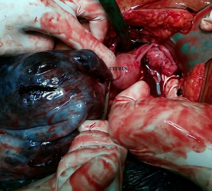
Intraoperatively 1.5 litres of hemoperitoneum was drained. There was evidence of left sided 12 x 9 cms size hemorrhagic ovarian cyst with torsion (Figure 1). A small rent of size 3 x 4 cm, with an active bleeder was found on the posterosuperior aspect of the cyst. The external surface of the cyst was smooth without any excrescences. Left fallopian tube was adherent to the cyst and was elongated and gangrenous due to simultaneous torsion. Uterus with right sided fallopian tube and right ovary were normal. Cyst detorsion was not done. A left sided salpingoophorectomy was performed. She received 3 units of packed cell transfusion in the perioperative period. She had an uneventful recovery and was discharged on the 5th post-operative day. Final histopathological diagnosis was a hemorrhagic ovarian cyst but with no viable ovarian parenchyma. There were no features of malignancy.


Figure1: Intraoperative photograph showing left sided large hemorrhagic ovarian cyst with normal right ovary and uterus. 

Discussion

Hemorrhagic ovarian cysts occur as a result of bleeding into the functional follicular and/ or corpus luteal cysts. Hence, they are almost always found in the women of reproductive age group and are nearly always benign. They may also be found in postmenopausal women who are on cyclic hormone replacement therapy. These cysts are usually small with an average diameter of 3.5 cm (range- 2.5- 8.5cm). [2] In our case, however, the cyst was unusually large with a diameter of 12 cm.
Patients may be asymptomatic or may present with acute pelvic pain, more commonly in the luteal phase of the menstrual cycle. The pain is attributed to the sudden intra-cystic hemorrhage leading to cyst enlargement and peritoneal irritation.[1] Cyst accidents such as torsion and rupture may also occur leading to signs and symptoms of acute abdomen, as was seen in our case. In such cases, other causes of acute abdomen such as ruptured ectopic pregnancy, endometriotic cyst rupture, pelvic inflammatory disease, acute appendicitis, mesenteric adenitis, Crohn’s disease etc need to be ruled out.[2]
Ultrasonography is the key imaging modality in the diagnosis of HOCs. The typical appearance is a thin walled cyst with smooth surface and an intra-cystic ‘Fishnet weave’ (reticular) pattern because of presence of interdigitating fibrin strands. A retracting blood clot within an anechoic cyst is also highly predictive of a HOC. Blood clots may mimic the solid component of an ovarian neoplasm, in which case a colour doppler ultrasonography should be advised. Absence of blood flow within the solid component suggests a blood clot rather than a neoplasm.[2]
Management of HOCs depends upon the clinical presentation, size of the cyst and ultrasound characteristics. Asymptomatic patients or those with mild symptoms, small cysts (usually <5 cm) and with a definitive ultrasound diagnosis, conservative treatment with analgesics, antibiotics and intravenous fluids is preferred. Spontaneous regression is known to occur within 2-6 weeks. Surgical management is indicated in symptomatic patients with larger cysts (> 5 cm), doubtful diagnosis and in those with signs/ symptoms of acute abdomen. 
In a study by Nair et al, conservative management of twisted HOCs in the form of detorsion and cystectomy was possible in 71.45% of patients. Most of them were young and in the early reproductive age group.  Approximately 15% patients required a radical surgery, all of whom were postmenopausal, in view of hemorrhagic necrosis of the adnexa. Thus, in our patient, owing to the age of the patient, completed family status, massive hemoperitoneum with rupture of the cyst, hemodynamic instability and extensive hemorrhagic necrosis of the adnexa, radical surgery in the form of unilateral salpingoohorectomy was performed. Detorsion was not performed as it would not have changed the management in this particular patient. A laparoscopic approach could be considered where resources and expertise are available.[1]

Conclusion

Hemorrhagic ovarian cysts are a common finding in routine clinical settings and are usually functional cysts with negligible malignant potential. In majority of cases, ultrasonography establishes the diagnosis owing to their characteristic appearance. Choice of conservative versus surgical management depends upon the clinical presentation and the size of the lesion, and emergency surgery may be mandated in a few. 

References
  1. Abbas AM, Amin MT, Tolba SM, Ali MK. Hemorrhagic ovarian cysts: Clinical and sonographic correlation with the management options. Middle East Fertil Soc J. 2016; 21(1): 41-5.
  2. Jain AK. Sonographic spectrum of hemorrhagic ovarian cysts. J Ultrasound Med. 2002; 21(8): 879-86.
  3. Nair S, Joy S, Nayar J. Five-year retrospective case series of adnexal torsion. J Clin Diagn Res. 2014;8(12): OC09-13.
Citation

Kale KG, Mali KA, Warke HS. Large hemorrhagic ovarian cyst with massive hemoperitoneum. JPGO 2018. Volume 5 No.2. Available from: http://www.jpgo.org/2018/02/large-hemorrhagic-ovarian-cyst-with.html

Abdominal Discharging Sinus Post Cesarean Section

Author Information

Saxena A*, Joshi AV**, Gupta AS***
(* First year Resident, ** Specialty Medical Officer, *** Professor.  Department of Obstetrics and Gynecology, Seth GS Medical College and KEM Hospital, Mumbai, India.)

Abstract

Sinus tracts are a common surgical condition. They may occur at various sites in the body. The common sites being pre-auricular, dental, perianal. Abdominal sinus tracts are generally rare. A sinus tract infection and its excision at the post cesarean section scar site has been reported here.

Introduction

Sinus tract infections of the abdomen are commonly caused either by deep infections or underlying suture materials. The deep infections likely to manifest as sinus tract are a ruptured appendicitis, recurrent gallstones, actinomycosis or deep seated pelvic infections.[1]

Case Report

A 30 year old, P2 L2 woman with previous 2 lower segment cesarean sections and tubal ligation done presented with complaints of purulent discharge and pain at suture site of the cesarean section. The last lower segment cesarean section was done 1 year back for fetal distress in first stage of labor through a vertical midline skin incision.The intrapartum and postpartum course was uneventful. She was discharged on day 4 postpartum. Suture removal was done on 12th post-operative day, and wound was healthy. She was asymptomatic until 2 months after surgery, when she noticed a small swelling at the lower end of the incision scar with discharge. The discharge was intermittent and non foul smelling. She had no complaints of fever or rash and no past medical chronic illness. She consulted a private practitioner for the same and was started on some medication. Her symptoms improved for a brief period of time, however it recurred on stopping the medication.
She then came to our hospital 8 months later. On examination, there was a small 0.5 x 0.5 cm swelling on the left side of the cesarean incision scar at the lower end. There were no signs of inflammation. However, there was some discharge through the opening over the swelling. A provisional diagnosis of sinus tract was made. Blood investigations that were done, were within normal limit. A wound swab of the discharge was sent which showed no growth.
An ultrasonography was done, which was suggestive of a 2 cc subcutaneous collection at incision site, an ultrasonography guided aspiration was attempted for the same but failed. She was given oral amoxycillin empirically for 5 days following which her symptoms improved for a brief period. A Computed Tomography (CT) scan with a sinogram was done on recurrence of the symptoms, which was suggestive of blind ending infra umbilical sinus tract with no intraperitoneal connection. A pus swab was sent for culture sensitivity, which was suggestive of Methicillin Sensitive Staphylococcus Aureus (MSSA) for which oral Linezolid 600 mg twice a day was started for 14 days as per the micro organism sensitivity report. The complaints resolved post antibiotic completion.
Two weeks later, she again complained of discharge through the scar site. A repeat ultrasound done showed a 3-4 ml collection at the local site in the subcutaneous plane with no deep communications.
A sinus tract excision was planned due to repeated recurrence of symptoms. Intra operative the sinus tract was found to be 3 cm deep with a horizontal extension noted on probing. The probe was kept and held in the tract to ensure entire excision of the tract. At the base of the tract was the suture knot of the previous surgery. The bulky knot of the suture material that was mono filament nylon was removed and the entire tract was excised by coring out. A wide excision was done en mass and sent for histopathology examination.
She came for follow-up two weeks later.  The wound was healthy and suture removal was done. There was no evidence of any remnant sinus. Histopathology report showed a fibrous tract with fibroblastic and lymphocytic infiltration that was consistent with a sinus tract.

Figure1. Sinus at the lower end of the midline vertical incision.

Figure 2. CT Sinogram

Figure 3. Excised entire sinus tract (arrow)

Discussion

Wound complications post cesarean section are a common entity. These maybe influenced by patient characteristics like obesity, anemia, malnutrition or certain procedure related characteristics like type of skin incision, any previous skin infections etc. The time of presentation can be immediate or late. The commonly encountered complications include surgical site infections, hematoma, wound dehiscence, scar granulomas, and sinus tract formation.
A sinus tract, often referred to as “tunneling wound”is a tract which is closed at one end. The major etiologies for this condition include infections, presence of a foreign body and tissue ischemia following trauma. The symptomatology can vary according to the site and etiology.
For infections presenting after a surgical procedure, foreign body is the most common cause.[2] Some patients may complain of recurring discharge from the scar site. They give a typical history of recurrent symptoms despite medical treatment.The cause of a persistent sinus discharge maybe the size or shape of the tract which leads to inadequate drainage of the cavity, thereby causing repeated bacterial proliferation.
The treatment is primarily surgical, which involves removing the retained foreign body like suture followed by a complete excision of the sinus tract. It is crucial to note the site, size and depth of the tract by probing. This ensures a complete assessment of the extent of the tract and will ensure a complete removal. Besides probing, a pre-operative sinogram can be done. However, it is vital to first obtain a wound swab to treat any underlying infections.
Braided suture materials pose a higher chance for wound infections.[3] However, in our patient, it was the retained mono filament nylon suture knot at the lower end of the rectus sheet that acted as a foreign body and nidus for infection causing the persistently discharging sinus tract. Nylon sutures are non-absorbable sutures and are commonly preferred for rectus sheath and skin approximation due to their high tensile property, permanent nature and minimal tissue inflammatory properties. This still caused a sinus tract which we presume to be secondary to wound infection that was harbored in the bulky knot. To completely cure the patient it is essential to excise the foreign body along with the entire sinus tract. This also prevents recurrence of the same.

Conclusion

Sinus tracts if not evaluated thoroughly can lead to persistent infections due to incomplete excisions. A comprehensive understanding of the wound etiology and complete assessment and excision of the extent of the tract is crucial in order to ensure a complete recovery.

References
  1. Surya M, Soni P, Nimkar K. Spontaneous Cholecysto-Cutaneous Fistula Draining Through an Old Abdominal Surgical Scar. Polish J Radiol. Medical Science International; 2016;81(10):498–501.
  2. Fajobi OA, Bari N, Mekwan J, Harrison C, Myint F. Chronic abdominal wall sinus and recurrent abscesses caused by a foreign body after laparotomy. J Wound Care 2005;14(9):427–8.
  3. Al-Mubarak L, Al-Haddab M. Cutaneous wound closure materials: an overview and update. J Cutan Aesthet Surg; 2013;6(4):178–88.
Citation

Saxena A, Joshi AV, Gupta AS. Abdominal Discharging Sinus Post Cesarean Section. JPGO 2018. Volume 5 No.2. Available from: http://www.jpgo.org/2018/02/abdominal-discharging-sinus-post.html